
Home Click on Images above to Enlarge
Home
Click on Images above to Enlarge
2008 IEEE International Ultrasonics Symposium (IUS)
Beijing International Convention Center (BICC)
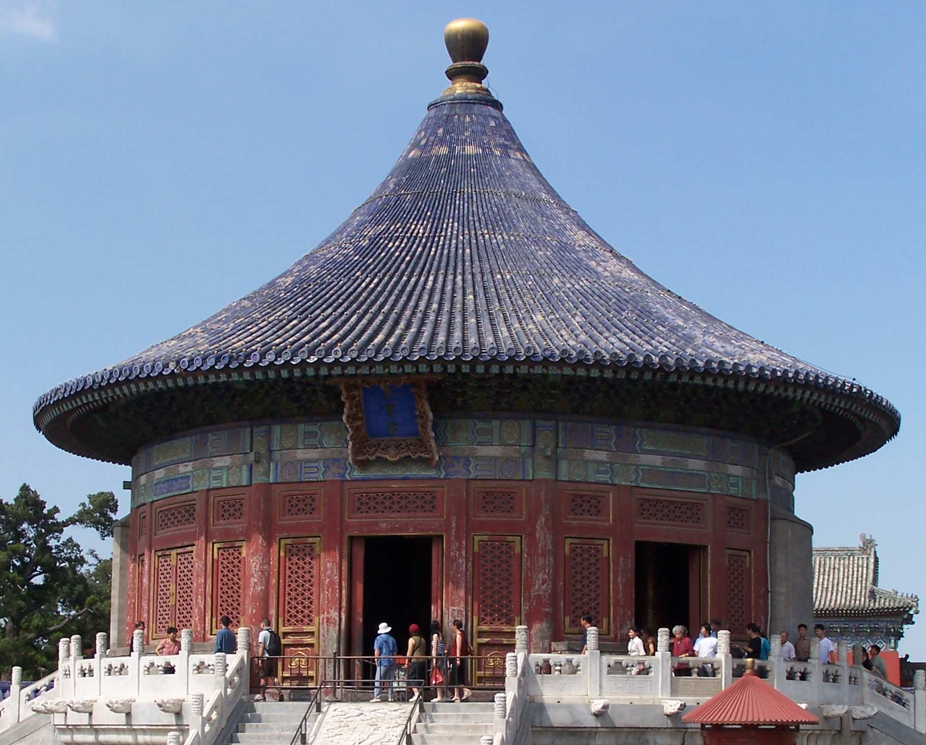
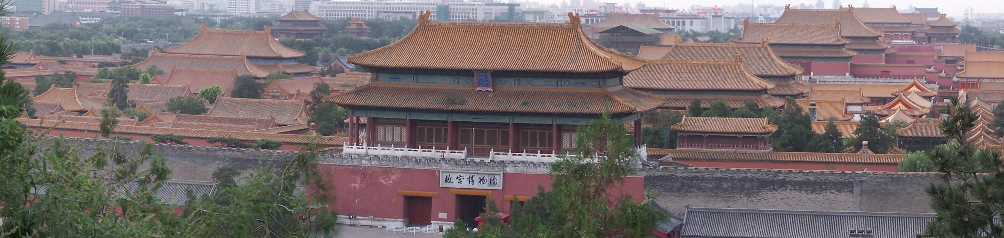
Beijing, China, November 2-5, 2008 (View: Conf. Photos/Videos and ![]() Beijing Photos
Beijing Photos ![]() )
)
|
|
Sponsored by the IEEE Ultrasonics, Ferroelectrics, and Frequency Control Society |
|
Student Paper Competition Winners (7 in Total)
Student Paper Competition Winners:
Seven winners of student paper competition were selected on Monday, November 3, 2008, from the 21 "Student Paper Competition Finalists" during the 2008 IEEE International Ultrasonics Symposium. The winners were also announced and honored during the Banquet and Shows of Tuesday evening, November 4, 2008.
Photos and videos of the ceremony of the winners of the student paper competition award are now available via "Conference Photos/Videos". The following is list of the award winners.
Finalist #1.1 (PS001-01) (3F-2):
Title: Design of Catheter for Combined Intravascular Photoacoustic and Ultrasound Imaging
Bo Wang (Presenter), Andrei Karpiouk, and Stanislav Emelianov, Biomedical Engineering, University of Texas at Austin, Austin, TX, USA. (Abstract ID: 309)
Abstract:
Background, Motivation and Objective:
Intravascular photoacoustic (IVPA) imaging is a promising imaging tool for detecting and differentiating the atherosclerotic plaques. Previously, we have demonstrated the utility of intravascular photoacoustic imaging using a laboratory system where the excised arterial tissue sample was irradiated with the laser beam from the outside while the intravascular ultrasound (IVUS) imaging catheter, inserted into the lumen, was used to receive the photoacoustic signal. However, for in-vivo IVUS and IVPA imaging, a combined catheter consisting of IVUS transducer and light delivery system is needed. In this paper we report our initial experience towards design and fabrication of a catheter capable of simultaneous IVPA and IVUS imaging.
Statement of Contribution/Methods:
The combined IVUS/IVPA imaging catheter was built based on a clinical, 40 MHz, single element IVUS catheter (Boston Scientific, Inc.). A 0.6 mm diameter optical fiber was used for light delivery. The proximal end of the fiber was coupled with a laser system. The distal tip of the optical fiber was polished at a 45 degree angle and placed inside a quartz tube. Both ends of the tube were sealed with epoxy to retain air around the fiber tip. As a result, such fiber assembly, when submersed into water, was irradiating the light sideway. The IVUS catheter was then attached to the optical fiber such that the ultrasound beam from the transducer and the laser beam from the optical fiber were aligned. To test the combined IVUS/IVPA imaging catheter, a model of the atherosclerotic vessel was fabricated. Specifically, within the otherwise homogeneous polyvinyl alcohol background, three 0.4% graphite inclusions of 1 mm diameter were positioned at different depths inside of the approximately 6-mm thick vessel wall to simulate various plaques in the artery. During imaging studies, the catheter was inserted into the lumen, and the phantom was rotated using a stepper motor. At each angular position, both photoacoustic and ultrasound A-lines were collected by a 14-bit GAGE A/D card operating at 200 MHz sampling rate. The IVUS and IVPA images were formed off-line from 256 equally spaced beams.
Results:
The IVPA images of the phantom obtained using the combined IVUS/IVPA catheter clearly identifies the inclusions located at specific depths. At the same time, co-registered IVUS images visualized the structure of the phantom. Finally, combined IVUS/IVPA images further outlined the location and extent of the inclusions within the vessel wall.
Discussion and Conclusions:
Overall, IVPA and IVUS images of sufficient quality were obtained using the initial prototype of the combined IVUS/IVPA catheter. Using optical fibers of smaller diameter, the size of the combined catheter can be further reduced. Therefore, our studies suggest that optical fibers can be used to deliver enough optical fluence for intravascular photoacoustic imaging of the vessel. Furthermore, other approaches in design of IVUS/IVPA imaging catheter will be discussed.
Finalist #1.4 (PS004-04) (2D-3):
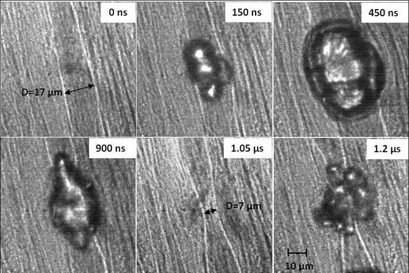
Title: Microbubble dynamics in microvessels: Observations of microvessel dilation, invagination and rupture
Hong Chen (Presenter), Andrew A. Brayman, Michael R. Bailey, and Thomas J. Matula, Center for Industrial and Medical Ultrasound, Applied Physics Laboratory, University of Washington, Seattle, WA, USA. (Abstract ID: 609)
Abstract:
Background, Motivation and Objective:
The fundamental interaction of an acoustically activated microbubble with small blood vessels is poorly understood. Understanding this interaction is important for designing better imaging schemes, and for targeting and drug delivery applications. High speed microscopy provides a tool to study interaction and response mechanisms.
Statement of Contribution/Methods:
Following an approved U.W. IACUC protocol, ultrasound contrast agent microbubbles, Evan’s blue and fluorescent dyes were perfused into the rat mesentery. These tissue samples were harvested for ex vivo observation. Evan’s blue was used to facilitate identification of microvessels and also as an indicator of blood vessel permeability changes. Fluorescence images were taken to examine the integrity of blood vessels. Tissue samples were exposed to short pulses of 1 MHz ultrasound. 14 high-speed microphotographic images were acquired for each experiment with shutter speeds of 50 ns and each image separated by 150 ns.
Results:
At low acoustic negative pressure (~1.5 MPa), bubble expansion caused microvessel dilation by approximately 1.2x. During bubble collapse, the vessel invaginated to approximately 0.9x of its original diameter (11 ?m). At high negative pressure (near 11 MPa), the vessel dilated by approximately 2.5x, followed by invagination of 0.4x of its original diameter (17 ?m). Vessel dilation and invagination were correlated temporally with bubble growth and collapse. At high pressure, the bubble and/or its fragments could be observed outside the original vessel, suggesting that the vessel had ruptured at some point. Vessel damage was also inferred by observation of fluorescent dye extravasation. An example of vessel dilation, invagination, and rupture can be seen in the following figure (pixel intensity values in the region around the blood vessel wall have been enhanced).
Discussion and Conclusions:
Our observations confirm some aspects of previous modeling and observational findings. However, direct observation of ultrasound-induced vessel invagination appears novel, and may be an important mechanism related to vessel damage. It remains uncertain if the vessel was damaged during dilation, invagination, or from a violent bubble collapse. It’s possible that both dilation and invagination contribute to vascular rupture. Work supported by NIH (5R01EB000350 and P01DK43881).
|
|
Finalist #1.8 (PS008-08) (1K-5):
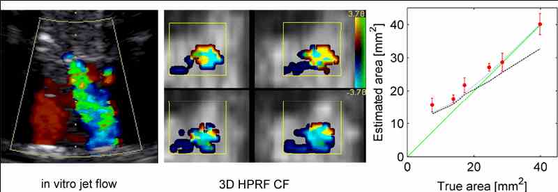
Title: Estimation of Valvular Regurgitation Area by 3D HPRF Doppler
*Torbjørn Hergum (Presenter), *Thomas Renhult Skaug, **Knut Matre, and Hans Torp, *Department of circulation and medical imaging, Norwegian University of Science and Technology, Trondheim, Norway, **Institute of Medicine, University of Bergen, Bergen, Norway. (Abstract ID: 1040)
Abstract:
Background, Motivation and Objective:
Determining the severity of leakage through a heart valve is important, but difficult. Two of the parameters which are clinically interesting in this regard are the area and the geometry of the lesion. Current practice for non-invasive measurement of the severity of valvular regurgitation is qualitative, and based upon using color flow- and spectral Doppler techniques.
Statement of Contribution/Methods:
In search for quantitative measurements of regurgitant severity we used 3D high pulse repetition frequency (HPRF) color flow imaging to measure the Doppler signal from multiple beams distributed over the laminar vena contracta region near the orifice. A steep clutter filter was used to separate the jet flow Doppler signals from the Doppler signals of the slowlymoving blood of the ambiguous sample volumes.
The power from the closely spaced ultrasound beams are summed to yield the total Doppler power, which is known to be proportional to the amount of blood moving above the clutter-filter cutoff velocity. The cross sectional area of the jet was found by scaling the summed Doppler power from these beams using both a-priori knowledge of the lateral extent of the beams and a reference beam which is completely covered by the orifice.
Both in vitro trials and computer simulation have been used for validation. The in vitro measurements were made using a pulsatile flow phantom holding porcine valves with six different holes, ranging from mild to severe mitral regurgitation. The method can be applied to other high-velocity valvular jets.
Results:
The mean value and standard deviation from the in vitro trials are plotted as red in the figure showing true area vs. estimated area. Two computer simulations are also included in the figure, the dashed and dotted lines are simulations respectively with- (blue) and without (black) stochastic variation.
Discussion and Conclusions:
Small holes of sizes comparable to a single ultrasound beam are overestimated as expected from simulations, and the estimates of the larger holes fits well with the line of identity (green). According to the stochastic simulations the method should underestimate the area of large orifices, but this is not seen in the in vitro data. Regardless of this the in vitro data enables us to distinguish between the different regurgitation degrees.
|
|
Finalist #2.2 (PS011-11) (5G-5):
Title: Improving the Bandwidth of Air Coupled Capacitive Ultrasonic Transducers Using Selective Networks
*Sean Mc Sweeney (Presenter) and **WMD Wright, *Electrical and Electronic Engineering Dept, University College Cork, National University of Ireland, Mallow, Cork, Ireland, **Electrical and Electronic Engineering, University College Cork, National University of Ireland, Cork, Cork, Ireland. (Abstract ID: 589)
Abstract:
Background, Motivation and Objective:
One of the key limitations on using CUT (Capacitive Ultrasonic Transducers) and cMUTs[1] (Capacitive Micromachined Ultrasonic Transducers) in air is their relatively narrow bandwidth which although superior to that of current piezoceramic devices[2] could be improved. Most air coupled capacitive devices could benefit hugely through the use of selective networks[3] for bandwidth expansion, resonance reinforcing, or a combination of both. This work has investigated the application of pole/zero manipulation techniques to modify and enhance the transmission characteristics of capacitive transducers through front end mounted components. The main objective was to positively enhance the performance characteristics of capacitive transducers.
Statement of Contribution/Methods:
A modified electrical equivalent circuit for CUTs to include the selective networks used was developed. The work assessed the effects of a tuned amplifier on the passband of the devices studied and then focused on more complicated network designs for enhancement. Simulations of the effects of the networks on the devices using equivalent circuit models were carried out and the response curves to pulsed operation were calculated and compared to experimental measurements from a pair of fixed CUTs with a combined centre frequency of 280kHz and 3dB bandwidth of 160kHz.
Results:
Increases in centre frequency of 25% and 3dB bandwidth of 77% using a single tuned amplifier were obtained. Resonance reinforcing, resonance shifting and ripple suppression were also studied through the manipulation of the q factor and pole location of such an amplifier. Performance enhancements were studied for a number of CUT aperture sizes and membrane thicknesses and a comparative study of the theoretical and experimental effect of these variations was conducted. With the appropriately designed network, enhancement of peak resonance with a simultaneous bandwidth expansion was obtained at the expense of other operating parameters such as stopband ripple. Simulations of more complicated circuit designs using equivalent circuit models of capacitive devices[4,5] showed that the maximum level of passband ripple observed for the bandwidth expansion method using a single tuned amplifier was reduced while achieving simultaneously the same 3dB results.
Discussion and Conclusions:
The implication for bandwidth expansion of a capacitive transducer through selective network design is significant, allowing increased resolution in imaging systems, ultrasonic ranging and non destructive evaluation. Significant improvements have been observed without additional signal manipulation, through digital means or otherwise, in certain transmission properties of the devices. Future work will expand on the enhancement of capacitive transducers through the use of hybrid resonator circuits and other related methods.
Finalist #3.2 (PS014-14) (6H-5):
Title: Towards thin film complete characterization using picosecond ultrasonics
Pierre-Adrien Mante (Presenter), Arnaud Devos, and Jean-François Robillard, IEMN-CNRS, France. (Abstract ID: 593)
Abstract:
Background, Motivation and Objective:
Mechanical characterization of thin films is a main issue in the microelectronic industry. The knowledge of these properties is necessary in many fields such as copper line interconnection and bulk acoustic wave resonators. A few techniques are reliable at this scale. Nano indentation or conventional laser-ultrasonic techniques can’t be effective in film thinner than 500 nm. Picosecond ultrasonics can also be used for thin film characterization. It is an efficient method to excite and detect vibrations within a thin film. A strong optical pulse warms a material surface, which leads to the creation of an acoustic wave propagating at the sound velocity. The waves propagation is longitudinal and it modifies the optical properties of the material. These modifications can be detected by a second time-shifted optical pulse.
Statement of Contribution/Methods:
In this technique we use a metallic very thin film as a transducer and only longitudinal waves can be generated. Due to that the full mechanical properties of thin layer cannot be measured. Here we show that thanks to a nanostructuration of the transducer, in-plane propagating waves are added using the same experimental setup. In the case of an isotropic medium, we have now access to all the acoustic properties.
Results:
We realized and studied 2D lattices of metallic nanocubes using e-beam lithography deposited onto the thin film to be charaterized. In a first experiment we will present results obtained on a 600nm-thick silica film.
Discussion and Conclusions:
Experiments were performed both on the lattices and out of the array of nanocubes. We respectively obtained the Rayleigh's velocity and the longitudinal velocity of silica. Then we can deduce Poisson's ratio and Young's modulus of silica: E=72GPa and nu=0.16, which is in very good agreement with literature. This first result demonstrates that we are able to extract longitudinal sound velocity, Rayleigh’s velocity, Young's modulus and Poisson's ratio in submicronic layers. Further results obtained on other materials isotropic and anisotropic will be also presented.
|
|
Finalist #4.3 (PS018-18) (4I-5):
Title: Shear mode BAW resonator based on c-axis oriented AlN thin film
Evgeny Milyutin (Presenter), and Paul Muralt, Ecole Polytechnique Federale de Lausanne, Switzerland. (Abstract ID: 522)
Abstract:
Background, Motivation and Objective:
Thin film bulk acoustic wave resonators (TFBAR's) also showed potential as gravimetric sensors. In contrast to RF filters working with longitudinal modes, bio-medical applications usually require detection in a liquid, thus must employ shear modes. The principle has recently been successfully demonstrated with TFBAR devices employing tilted c-axis growth of ZnO [1, 2]. In this work, we show that it is also possible to use non-tilted AlN thin films when interdigitated (ID) electrodes (IDE) are used. A true shear BAW thickness mode can be excited. Parasitic Lamb waves are avoided by the use of acoustic reflectors.
Statement of Contribution/Methods:
Performance and design of shear modes in AlN(001) films excited by ID electrodes were simulated by finite element modeling using the boundary element method (FEM-BEM). Devices have been fabricated with 1.5 microns thick (001)-textured AlN thin films on top of a Bragg reflector composed of 5 double layers of SiO2/AlN. The Al electrode system was defined by photolithography along with a lift-off process.
Results:
The performances of resonators were assessed in air and silicon oil. Typically resonance frequency of the devices was between 1.8-1.9GHz. By using different electrode periodicities, the BAW nature of the resonance was confirmed through the absence of a shift. A quality factor of about 1000 was achieved when operated in air. Under immersion, the Q-factor decreased to 260. Experimental results are in a good agreement with simulations, when we consider acoustic emission through the Bragg grating as the only loss factor.
Discussion and Conclusions:
The achieved results and the simplicity of fabrication of proposed device show their potential as gravimetric sensors for immersed applications. The achieve Q-factor is higher than reported in literature for tilted c-axis resonators [3]. Further optimization of design and materials is going on. The integration of an immobilization layer is in development.
1. Link, M., M. Schreiter, J. Weber, R. Gabl, D. Pitzer, R. Primig, W. Wersing, M.B. Assouar, and O. Elmazria, Caxis inclined ZnO films for shear-wave transducers deposited by reactive sputtering using an additional blind.
J.Vac.Sci.Techn. A, 2006. 24: p. 218-222.
2. Weber, J., W.M. Albers, J. Tuppurainen, M. Link, R. Gabl, W. Wersing, and M. Schreiter, Shear mode FBAR as highly sensitive liquid biosensors. Sensors and Actuators A, 2006. 128: p. 84-88.
3. G. Wingqvist, J. Bjurstrom, L. Liljeholm, V. Yantchev, I. Katardjiev, Shear mode AlN thin film electro-acoustic resonant sensor operation in viscous media, Sensors and Actuators B 123 (2007), 466-473.
Finalist #5.1 (PS019-19) (4D-3):
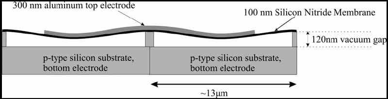
Title: Investigation of charge diffusion in Capacitive Micromachined Ultrasonic Transducers (CMUTs) using optical interferometry
Hanne Martinussen (Presenter), Astrid Aksnes, and Helge E. Engan, Electronics and Telecommunications, Norwegian University of Science and Technology, Trondheim, Norway. (Abstract ID: 274)
Abstract:
Background, Motivation and Objective:
Capacitive Micromachined Ultrasonic Transducers (CMUTs) have been developed and fabricated at our department. The main goal is to use an improved version of these structures to perform medical imaging to detect unstable plaque in the coronary arteries. Unstable plaques are fatty lipid pools contained in the wall of the coronary arteries by a thin fibrous cap. A rupture of this cap can lead to an infarction. The CMUTs have a radius of 5.7?m and a center frequency of about 30MHz in air. When an RF voltage is applied in addition to a DC bias the membrane will vibrate and generate ultrasound waves. This DC bias is in the order of 30V and leads to a charge diffusion in the CMUTs. This work investigates this process in detail.
Statement of Contribution/Methods:
A heterodyne interferometer has been built in order to characterize the CMUTs. The setup can measure absolute phase and amplitudes. By using two acousto-optic modulators in the reference arm of the interferometer we can measure acoustic frequencies in the range 10kHz-1.2GHz. The results from the interferometer are supplemented with measurements from a network analyzer. The network analyzer takes the mean of all currents generated by CMUTs whereas the interferometer inspects individual CMUT elements.
Results:
The vibrating membrane in the CMUT is made of silicon nitride, which ideally is an insulator. However, we observe a charge diffusion through this membrane influencing the response of the CMUTs. There are two possible mechanisms. One is that positive charges diffuse from the bottom electrode through the silicon substrate and into the silicon nitride membrane. The other is that negative charges from the top electrode diffuses into the silicon nitride membrane. An experiment investigating the resonance frequency as a function of time indicated that the latter mechanism is dominant. Measurements from both the interferometer and the network analyzer supported this conclusion.
Discussion and Conclusions:
The measurements presented here are performed in air. Under loading conditions such as water or tissue the frequency bandwidth of the CMUT increases substantially. The charge diffusion problem may therefore not be a major problem when the CMUT is operated in water.
|
|
Home Contact the webmaster, Dr. Jian-yu Lu, for questions. © Copyright 2006-2008 IEEE UFFC Society