
Home Click on Images above to Enlarge
Home
Click on Images above to Enlarge
2008 IEEE International Ultrasonics Symposium (IUS)
Beijing International Convention Center (BICC)
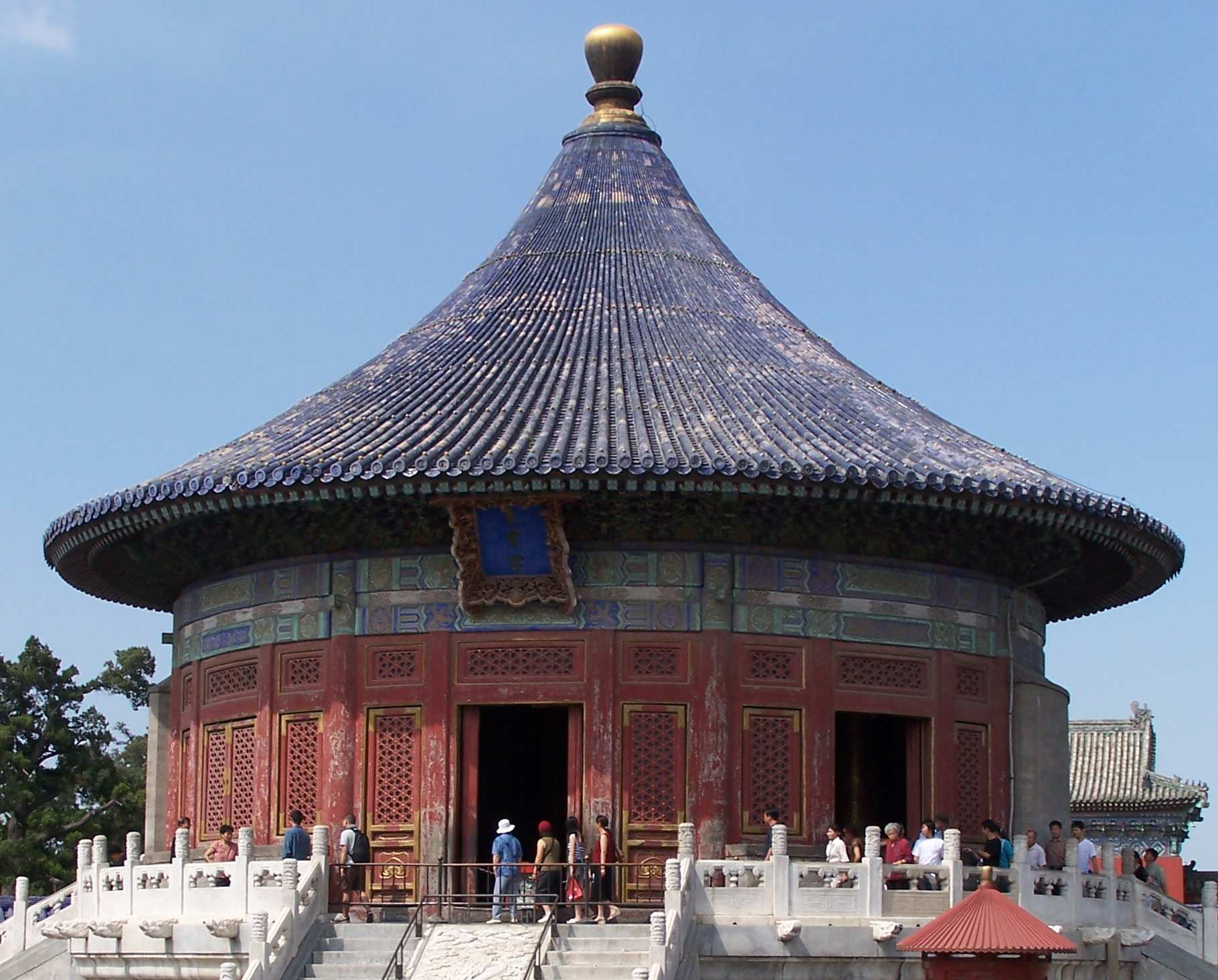
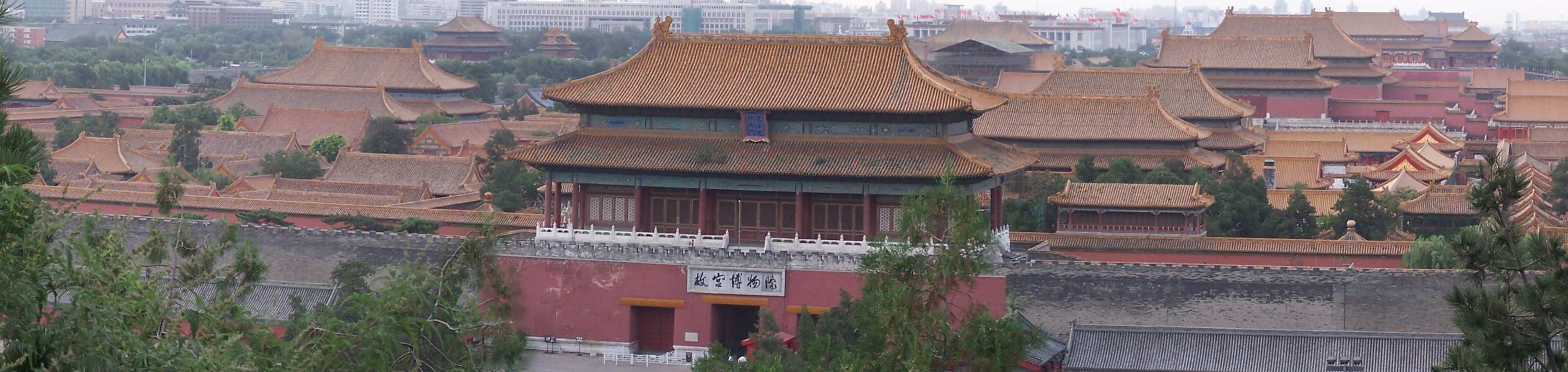
Beijing, China, November 2-5, 2008 (View: Conf. Photos/Videos and ![]() Beijing Photos
Beijing Photos ![]() )
)
|
|
Sponsored by the IEEE Ultrasonics, Ferroelectrics, and Frequency Control Society |
|
Short Courses (12 in Total)
Overview of Short Courses (Please Click on the Links to Jump to the Courses):
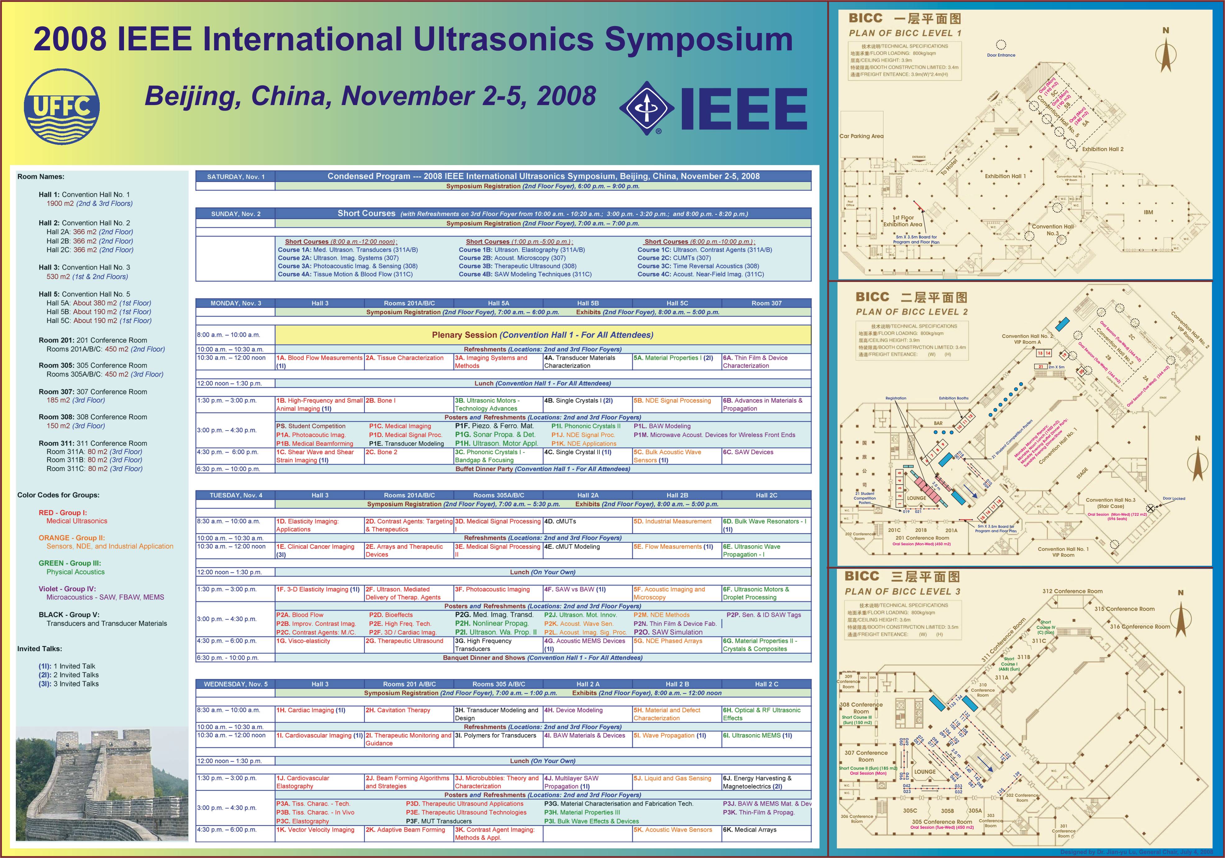
Notes: To find the short course rooms and their locations, please check the Condensed Program and the Floor Plan. Detailed technical program of the conference can be found in the Full Program (Program Book), Abstract Book, and Meeting Planner.Short Course Evaluation Form:
If you are going to attend the short course(s), please download the Short Course Evaluation Form below, fill it out after each short course, and then return it to the conference registration desk to help us to improve the courses in the future.|
Short Course Evaluation Form for Short Course Attendees: |
Short Course 1A (8:00 A.M. - 12:00 Noon, Sunday, November 2, 2008):
Course Title: Medical Ultrasound Transducers
Douglas G. Wildes and L. Scott Smith, GE Global Research Center, Niskayuna, NY, USA.
Course Description: This course will provide an introduction to the design, fabrication, and testing of medical ultrasound transducers. Starting from an overview of the basic types of phased-array transducers (linear, convex, sector), we will discuss how the design for a probe is derived from its target application and how equivalent-circuit, finite-element, and acoustic field models can be used to optimize the design and accurately predict performance. A discussion of the structure of an ultrasound probe will lead to a survey of the different types of materials used in probes and their critical properties. Typical fabrication processes will be introduced and common problems in probe manufacturing will be summarized. Methods for evaluating completed transducers will be discussed. The course will highlight recent developments in probe technology, including single crystal piezoelectrics, cMUT transducers, catheters, multi-row and 2D arrays, and electronics in probes, and will discuss performance advantages and fabrication difficulties which may be associated with each.
Douglas G. Wildes is a physicist with GE Global Research. He earned an A.B. in physics and mathematics from Dartmouth College and a Ph.D. in low-temperature physics from Cornell University, then joined GE in 1985. Since 1991, Dr. Wildes?research has focused on aperture design, fabrication processes, and high-density interconnect technology for multi-row and 4D imaging transducers for medical ultrasound. Dr. Wildes has 23 issued patents and 19 external publications. He is a member of the American Physical Society and a Senior Member of the IEEE.
L. Scott Smith is a physicist with GE Global Research. He earned B.S. and Ph.D. degrees in physics from the University of Rochester and the University of Pennsylvania respectively. Joining GE in 1976, he developed phased array probes for medical ultrasound. More recently, he examined novel probe materials and led projects on pediatric endoscopes and adaptive acoustics. Dr. Smith has 43 issued patents and over 35 refereed publications. He is a member of the American Physical Society and a Senior Member of the IEEE where he serves as Vice Chair for Transducers on the Ultrasonics Symposium’s Technical Program Committee.
Short Course 2A (8:00 A.M. - 12:00 Noon, Sunday, November 2, 2008):
Course Title: Ultrasound Imaging Systems: from Principles to Implementation
Kai E. Thomenius, GE Global Research Center, Niskayuna, NY, USA.
Course Description: The design of medical ultrasound imagers is undergoing important changes brought about by advances in semiconductors and signal/image procession technologies coupled with changes in medical practice and the utilization of medical imaging in general. Unique aspects of data acquisition and processing in the ultrasound scanner enable opportunities not available to other imaging modalities. The goal of this course is to review the system design of ultrasound scanners from a linear systems point of view including transduction, beam formation, and image formation functions. We will discuss analytical methods used in developing the design of a scanner in use today. The key points to be covered deal with methods of analysis of array data, the interaction of transmit and receive beams with clinically relevant targets, and how this interaction is used in the generation of clinically useful images. The means by which these analytical methods contribute to a system design and the trade-offs involved are reviewed. The last several years have seen steady migration of functionality into software; this has enabled significant miniaturization of scanners. The impact of this on system design and the size of ultrasound scanners of the future will be discussed.
Kai E. Thomenius is a Chief Technologist in the Imaging Technologies Organization at General Electric’s Global Research facility in Niskayuna, NY, USA. His focus is on Ultrasound and Biomedical Engineering. Previously, he has held senior R&D roles at ATL Ultrasound Inc., Interspec Inc., Elscint Inc., as well as several other ultrasound companies. In addition, he is currently an Adjunct Professor in the Electrical, Computer, and Systems Engineering Department at Rensselaer Polytechnic Institute where he teaches a course in general imaging. Dr. Thomenius?academic background is in electrical engineering with a minor in physiology; all of his degrees are from Rutgers University. His long-term interests have been in ultrasound beam formation and miniaturization of ultrasound scanners, propagation of acoustic waves in inhomogeneous media, and determination of physiological information from the echoes that arise from such beams. Dr. Thomenius is a Fellow of the American Institute of Ultrasound in Medicine.
Short Course 3A (8:00 A.M. - 12:00 Noon, Sunday, November 2, 2008):
Course Title: Photoacoustic Imaging and Sensing
Stanislav Emelianov, Biomedical Engineering Department, University of Texas at Austin, USA.
Course Description: This course is designed to provide both a broad overview and a comprehensive understanding of photoacoustic (also known as optoacoustic and, more generally, thermoacoustic) imaging, sensing and spectroscopy. With a brief historical introduction, we will begin the course by examining the foundations of photoacoustics, including derivations and a discussion of governing equations. We will also review relevant optical properties of the tissues and related topics of laser-tissue interaction. The experimental aspects of photoacoustic imagining and sensing will then be discussed with emphasis on system hardware and signal/image processing algorithms. Techniques to increase contrast and to differentiate various tissues in photoacoustic imaging will be presented. The course will conclude with an overview of several experimental systems capable of photoacoustic imaging, and discussion of current and potential biomedical and clinical applications of photoacoustics.
Stanislav Emelianov received B.S. and M.S. degrees in Physics and Acoustics in 1986 and 1989, respectively, from the Moscow State University, and a Ph.D. degree in Physics in 1993 from the Moscow State University and the Institute of Mathematical Problems of Biology of the Russian Academy of Science. In 1989, he joined the Institute of Mathematical Problems of Biology, where he was engaged in both mathematical modeling of soft tissue biomechanics and experimental studies of noninvasive visualization of the mechanical properties of tissue. Following his graduate work, he moved to the University of Michigan, Ann Arbor, as a post-Doctoral Fellow in the Bioengineering Program and in the Electrical Engineering and Computer Science Department. From 1996 to 2002, Dr. Emelianov was a Research Scientist at the Biomedical Ultrasonics Laboratory of the Biomedical Engineering Department at the University of Michigan. During his tenure at Michigan, Dr. Emelianov was involved primarily in the theoretical and practical aspects of elasticity imaging using ultrasound and MRI. Dr. Emelianov is currently teaching and conducting research in the Department of Biomedical Engineering at the University of Texas at Austin. His research interests are in medical imaging and therapeutics, including ultrasound, photoacoustic, elasticity and multi-modality imaging, photothermal therapy, cellular/molecular imaging and therapy, functional imaging, etc.
Short Course 4A (8:00 A.M. - 12:00 Noon, Sunday, November 2, 2008):
Course Title: Estimation and Imaging of Tissue Motion and Blood Velocity
Hans Torp and Lasse Lovstakken, Department of circulation and medical imaging, Norwegian University of Science and Technology, Trondheim, Norway.
Course Description: This course provides a basic understanding of the physical principles and signal processing methods for estimation of blood and tissue motion. The course begins with an overview of currently used techniques for velocity estimation using pulsed- and continuous-wave Doppler, and color flow imaging. Statistical models for the received signal, as well as commonly used velocity estimators will be developed. Simulation methods for ultrasound signals from moving blood and tissue will be discussed and examples in Matlab will be shown. The suppression of clutter from slowly moving targets is central to all processing schemes and will be given special attention. Also, current methods of tissue velocity and strain rate imaging will be given special elaboration. More advanced topics will also be covered. An overview of current adaptive filter schemes for attenuating clutter will be given, and 2-D / 3-D vector velocity estimation techniques will also be presented. The principles and practical limitations of these methods will be discussed, and potential applications in blood velocity imaging and myocardial velocity- and strain imaging will be shown.
Hans Torp received the MS degree in mathematics in 1978, and the Dr. Techn. Degree in electrical engineering in 1992; both from the University of Trondheim, Norway. Since 1980 he has been working with ultrasound technology applied to blood flow measurements and imaging at the University of Trondheim, in cooperation with GE-Vingmed Ultrasound. He is currently professor of medical technology at the Norwegian Univesity of Science and Technology, and has since 1987 given courses on ultrasound imaging and blood flow measurements for students in electrical engineering and biophysics. His research interests include statistical signal- and image processing with application in medical ultrasound imaging.
Lasse Lovstakken received the Masters degree in Engineering Cybernetics in 2002 and a PhD in Medical Technology in 2007, both at the Norwegian University of Science and Technology, in Trondheim, Norway. He is currently working as a post doctoral research fellow at the Department of Circulation and Medical Imaging at the Norwegian University of science and Technology. His research interests include signal and image processing with applications in ultrasound imaging, with a special focus on imaging of blood and tissue movement.
Short Course 1B (1:00 P.M. - 5:00 P.M., Sunday, November 2, 2008):
Course Title: Ultrasound Elastography: Quantitative Approaches
*Jeffrey Bamber and **Paul Barbone, *Institute of Cancer Research and Royal Marsden Hospital, UK. **Boston University, USA.
Course Description: There is evidence that ancient cultures extending back thousands of years used palpation to assess the mechanical properties of tissues, and thus detect and characterise disease or injury. Simple palpation continues to be of value in modern medicine, both practiced by doctors and as a technique for self-examination, but palpation is limited to a few accessible tissues and organs, and the interpretation of the information sensed by the fingers is highly subjective. Ultrasound elastography aims to display images that are related to a broad range of parameters that describe the spatial and temporal variations in tissue viscoelasticity. It does so by processing time-varying echo data to extract the spatial and/or temporal variation of a stress-induced tissue displacement or strain. In recent years the method early form has emerged as a real-time imaging modality available as an option on several commercial ultrasound systems, and is starting to prove clinically valuable, for example in breast cancer diagnosis. Nevertheless, in its present form it remains a strongly subjective technique and continues, as with palpation, to require considerable interpretive skills to be learnt. There are good reasons to believe that a more quantitative and objective analysis will lead to clinically more valuable measures of tissue composition, function or state, with images that are easier to interpret. This short course will outline some of the limitations and pitfalls of current elastographic methods, and will then introduce the opportunities for, potential value of and challenges for making elastography more quantitative. It will then review work on modeling tissues and their mechanical behavior, the fundamentals of ultrasound elastographic experimental techniques required for quantitative imaging, the use of static, vibrational and impulsive loads, the inverse methods for measurement and image reconstruction, methods for stress measurement, and shear wave propagation methods. This will lead to a discussion of the likely consequences for medical applications and future instrumentation. Examples of results will be presented for a range of medical application areas and for various mechanical characteristics such as shear modulus, nonlinearity, anisotropy, friction at mechanical discontinuities, as well as properties that determine viscoelastic and poroelastic behavior.
Jeffrey Bamber is head of the Ultrasound and Optics Physics Team, and is Senior Tutor for the Research Degrees Program at The Institute of Cancer Research Sutton, U.K. He has an honorary position as a Medical Physicist within the Royal Marsden Hospital, Sutton. He received a BSc in Physics from the University of Kent at Canterbury in 1972, an MSc in Biophysics and Bioengineering from the University of London in 1974, and a PhD in Biophysics in 1980, also from the University London. He continued as a research scientist following his PhD at the Institute of Cancer Research, becoming a team leader in 1986. His research interests have included: acoustic characteristics of tissues, ultrasound image speckle and texture, speckle reduction, ultrasound aberration, psychophysics of perception of information in ultrasound images and movies, ultrasonic methods in breast cancer, measurement of tumor volume and blood flow, ultrasound tissue motion tracking, tissue elasticity imaging, temperature imaging, high frequency ultrasonic imaging and tissue characterization, ultrasound and optical methods in skin cancer, microbubble contrast agents, ultrasound guidance of focused ultrasound therapy and radiotherapy, ultrasound in radiation dosimetry, microbubbles as gene therapy vectors, and molecular imaging. Prizes for work to which he has contributed include 5 best paper awards in peer reviewed journals and 2 book publishing awards for excellence. He is a past vice-president of the International Society for Skin Imaging, a past president of the International Association for Breast Ultrasound, and currently serves on the Council of the British Medical Ultrasound Society.
Paul E. Barbone is Associate Professor of Mechanical Engineering at Boston University. He received Bachelors of Engineering Science and Mechanics from Georgia Institute of Technology in 1986, a Masters of Mechanical Engineering in 1987 from Stanford University, and a PhD in Mechanical Engineering from Stanford University in 1992. He did postdoctoral research at the University of Cambridge (1992-1993) in the Department of Applied Mathematics and Theoretical Physics, served as lecturer at School for Advanced Studies in Industrial and Applied Mathematics, Valenzano, Italy (1992), and was Haddow Fellow and visiting Researcher at the Institute of Cancer Research, Sutton, UK (2000-2001). His research approach is mathematical and theoretical analysis. He works mainly on forward and inverse problems in acoustics and solid mechanics, and sidelines in the analysis of computational formulations. Over the past several years, his research focus has been inverse problems in "Biomechanical Imaging:" imaging the mechanical properties of tissues in situ and in vivo. His research work has been recognized through prizes from US National Science Foundation, US Office of Naval Research, Acoustical Society of America, and the J. William Fulbright Foundation.
Short Course 2B (1:00 P.M. - 5:00 P.M., Sunday, November 2, 2008):
Course Title: Acoustic Microscopy - Fundamentals and Applications
*Roman Gr. Maev, **Naohiro Hozumi, ***Kazuto Kobayashi, and ****Yoshifumi Saijo, *Centre for Imaging Research and Advanced Materials Characterization, University of Windsor, Ontario, Canada. **Department of Electrical & Electronic Engineering, Aichi Institute of Technology, Toyota, Japan. ***Honda Electronics Co. Ltd., Aichi, Japan. ****Tohoku University, Sendai, Japan.
Course Description: The goal of this course is to introduce the fundamentals and major principles of scanning acoustic microscopy. This course aims to describe advanced acoustic microscopy methods for investigating the microstructure and physical mechanical properties of materials of different nature, from crystalline to biomaterials. The materials discussed during this course cover most aspects of physical principles and applications of high-resolution acoustic microscopy and reflects the modern research status in this field. Included are different topics in physical acoustics, ultrasound, solid state physics, materials characterization and nondestructive evaluation. Special attention will be paid to the principle and application of several types of scanning acoustic microscopes for medical and biological use. Progress in digital measurement and pulse technology has remarkably upgraded the performance of these types of microscopes and this will be described within the course. The sound speed microscope which conventionally used tone-burst and analog phase detector was improved in accuracy, stability and operation ability. It can be used for characterization of tissue sliced and mounted on a slide glass. It can visualize not only acoustic impedance but bulk modulus, attenuation constant and density. The acoustic impedance microscope can visualize the acoustic impedance of a cross section in touch with a plastic substrate by transmitting an acoustic beam from the rear side of the substrate. This type of microscopy has an advantage that the measurement can be performed in vivo, introducing no contamination into the target system. With a wide frequency range up to 400 MHz, both types of microscopes can observe with a special resolution as fine as cell structure. Discussed will be the principle of the sound speed and acoustic microscopes driven by a wide band pulse and several examples of observation of cerebella tissue and cultured cells will be shown. In addition, there will be a presentation of recent results in acoustic microscopy technology development achieved by Honda Electronics (Japan) and Tessonics (Canada). The detail of the hardware and software of those microscopes that are commercially available will be described. The prototype microscopes have been improved a lot after being commercialized. The hardware, software and biomedical applications of these microscopes will be described with a large number of examples as additional illustrations. This course will conclude with an overview of the future perspectives of the general principles of microscopic observation using various ultrasound waves as well as the most promising future applications.
Roman Gr. Maev received his Ph.D. from the Physical Institute of the Russian Academy of Sciences in 1973 and his D.Sc. in acoustic microscopy from the Russian Academy of Sciences, Moscow, in 2002. From 1994 to 1997, he held a post as Director of the Acoustic Microscopy Center of the Russian Academy of Sciences, then established a Centre for Imaging Research and Advanced Material Characterization at the University of Windsor, Canada. He is currently a Full Faculty Professor at the Physics Department of the same University and since 2001 the Chairholder of the NSERC/DaimlerChrysler/Industrial Research Chair in Applied Solid State Physics and Material Characterization. Professor Maev’s research interests focus on the fundamentals of condensed matter, physical acoustics, ultrasonic imaging, and acoustic microscopy. He has published numerous books, more than 300 scientific papers, and holds twenty patents.
Naohiro Hozumi was born in Kyoto, Japan on April 2, 1957. He received his B.S., M.S. and Ph.D. degrees in 1981, 1983 and 1990 from Waseda University. He was engaged in Central Research Institute of Electric Power Industry (CRIEPI) from 1983 to 1999. He was an associate professor of Toyohashi University of Technology from 1999 to 2006. Since 2006, he has been a professor of Aichi Institute of Technology. He has been engaged in the research in insulating materials and diagnosis for high voltage equipment, acoustic measurement for biological and medical applications, etc. He was awarded in 1990 and 1999 from IEE of Japan for his outstanding research papers. He is a member of IEEE, IEE of Japan and the Acoustic Society of Japan.
Kazuto Kobayashi was born in Aichi, Japan on June 8, 1952. He received B.S. degree in electrical engineering from Shibaura Institute of Technology, Tokyo, Japan in 1976. He is currently a director of Department of Research and Development at Honda Electronics Co. Ltd. in Toyohashi, Japan. His research activities and interests include medical ultrasound imaging, signal processing and high frequency ultrasound transducers.
Yoshifumi Saijo was born in Yokohama, Japan on July 21, 1962. He received the M.D. and the Ph.D. degrees in 1988 and 1993 from Tohoku University. He is currently a Professor of the Department of Biomedical Imaging at the Graduate School of Biomedical Engineering of Tohoku University. He is concurrent with Institute for International Advanced interdisciplinary Research of Tohoku University and the Department of Cardiovascular Surgery of Tohoku University Hospital. His main research interests are assessment of biomechanics of cells and tissues by high frequency ultrasound and clinical ultrasonic evaluation of cardiovascular system with intravascular ultrasound and transesophageal echocardiography. He was awarded in 1997 for his outstanding research paper in Ultrasound in Medicine and Biology, the official journal of the World Federation of Ultrasound in Medicine and Biology. He is a member of The Japan Society of Ultrasonics in Medicine, Japanese Society of Echocardiography and Japan Circulation Society.
Short Course 3B (1:00 P.M. - 5:00 P.M., Sunday, November 2, 2008):
Course Title: Therapeutic Ultrasound
Lawrence A. Crum, Applied Physics Laboratory, University of Washington, Seattle, WA, USA.
Course Description: The use of ultrasound in medicine is now quite commonplace, especially with the recent introduction of small, portable and relatively inexpensive, hand-held diagnostic imaging devices. Moreover, ultrasound has expanded beyond the imaging realm, with methods and applications extending to novel therapeutic and surgical uses. These applications broadly include: Tissue ablation, acoustocautery, body contouring, site-specific and ultrasound mediated drug activity, extracorporeal lithotripsy, and the enhancement of natural physiological functions such as wound healing and tissue regeneration. A particularly attractive aspect of this technology is that diagnostic and therapeutic systems can be combined to produce totally non-invasive, image-guided therapy. This general lecture will review a number of these exciting new applications of ultrasound and address some of the basic scientific questions and future challenges in developing these methods and technologies for general use in our society. We shall particularly emphasize the use of High Intensity Focused Ultrasound (HIFU) in the treatment of benign and malignant tumors as well as the introduction of acoustic hemostasis, especially in organs which are difficult to treat using conventional medical and surgical techniques.
Lawrence A. Crum is currently Principal Physicist in the Applied Physics Laboratory and Research Professor of Bioengineering and Electrical Engineering at the University of Washington. He has held previous positions at Harvard University, the U. S. Naval Academy and the University of Mississippi, where he was F. A. P. Barnard Distinguished Professor of Physics and Director of the National Center for Physical Acoustics. He has published over 300 articles in professional journals, holds an honorary doctorate from the Universite Libre de Bruxelles, and was recently awarded the Helmholtz-Rayleigh Silver Medal of the Acoustical Society of America. He is Past President of the Acoustical Society of America, the World Council on Ultrasonics, and of the Board of the International Commission for Acoustics.
Short Course 4B (1:00 P.M. - 5:00 P.M., Sunday, November 2, 2008):
Course Title: SAW Modelling Techniques
Victor P. Plessky, GVR Trade SA, Bevaix, Switzerland.
Course Description: This course provides introduction to the design techniques of SAW devices. The course includes and will discuss: a) SAW excitation on piezoelectrics by linear charges, elementary theory of the Interdigital Transducer (IDT) with non-reflecting electrodes, design of typical IDTs on quartz and LiNb, delay lines characteristics and matching issues. b) Single Phase Unidirectional Transducer (SPUDT)- design and applications. c) Propagation of SAW in periodic structures, coupling of modes?(COM) model, and simulation with COM model of IDTs and reflectors. d) Modeling of SAW devices based on Green’s function software. e) CRF/DMS filter design ?examples of?device simulation; optimization software f) Synchronous resonators, extraction of COM parameters, and ladder filters design. g) Design of SAW-tags. During the lecture, the attendee will see demonstrations of design processes for typical filter specifications. The COM model will be presented in details sufficient for practical use. The course will conclude with a review of unsolved problems and challenges in the SAW devices design area.
Victor P. Plessky was born near Gomel, Belarus.?He now lives and works in Switzerland. Before leaving the USSR in 1991, he worked as a head of laboratory in IRE of Academy of Sciences in Moscow region in Russia. He received his Ph.D. degree from the Moscow Institute of Physics and Technology in 1978,?and received his Doctor of Science degree in physics and mathematics from the Institute of Radio-engineering and Electronics (IRE RAS, 1987). He received the Full Professor title from the Russian Government in1995. For the last 16 years he has worked in Switzerland, first as a Principal Scientist at the company Micronas SA. He now is an owner and CEO of the consulting company GVR Trade SA. His main spheres of interest are theory of microacoustics, surface acoustic waves (SAW) theory and devices, devices for signal filtering and frequency control, SAW sensors and SAW-tags.?A few of his works in periodic structures have received wide recognition. Dr. V. Plessky worked as Visiting Professor in HUT (Finland), Freiburg University (Germany), Uppsala University (Sweden), EPFL (Switzerland). He has authored or co-authored over 200 papers and many patents. For many years he serves ad TPC member of the IEEE Ultrasonics Symposium.
Short Course 1C (6:00 P.M. - 10:00 P.M., Sunday, November 2, 2008):
Course Title: Ultrasound Contrast Agents: Theory and Experiment
*Nico de Jong and **Michel Versluis, *Erasmus MC, The Netherlands. **University of Twente, The Netherlands.
Course Description: The course consists of 6 topics: a) An overview will be presented of the (clinical and pre-clinical available) contrast agents, including the properties and characteristics of the gas inside the bubble and the shell surrounding it. b) Models of the behavior of small bubbles in an ultrasound field will be discussed. Simple models based on a one dimensional mass-spring system and more complicated models including gas and shell properties. c) Experimental acoustic methods for UCA will be presented for characterizing the bubbles in suspension, including harmonic and sub-harmonic scattering, absorption and attenuation. Also the influence of ambient pressure, temperature and gas concentration will be discussed. d) Experimental optical and acoustical methods for characterizing individual bubbles. e) Imaging methods for contrast agents, e.g. fundamentals, harmonic, subharmonic and superharmonic and multi-pulse methods like the pulse inversion, power modulation etc. and new methods including chirp excitation and radical modulation. f) Molecular imaging and ultrasound mediated drug delivery: Interaction between mammalian cells and ultrasound in the presence of (targeted) bubbles will be discussed.
Nico de Jong graduated from Delft University of Technology, The Netherlands, in 1978. He got his M.Sc. in the field of pattern recognition. Since 1980, he has been a staff member of the Thoraxcenter of the Erasmus University Medical Center, Rotterdam, The Netherlands. At the Dept. of Biomedical Engineering, he developed linear and phased array ultrasonic probes for medical diagnosis, especially compound and transesophageal transducers. In 1986 his interest in ultrasound applications shifted toward the theoretical and practical background of ultrasound contrast agents. In 1993 he received his Ph.D. for “Acoustic properties of ultrasound contrast agents.? His current interests are 3D (matrix) transducers, bubble behavior and fast framing camera systems. Since 1996 he organizes, together with the cardiologist Dr. Folkert ten Cate, the annual European Symposium on Ultrasound Contrast Imaging, held in Rotterdam and attended by approximately 175 scientists from all over the world. Since 2003 Nico de Jong is part-time professor at the University of Twente.
Michel Versluis graduated in Physics in 1988 at the University of Nijmegen, the Netherlands, with a special interest in Molecular Physics and Astrophysics. Later, he specialized in the application of intense tunable UV lasers for flame diagnostics resulting in a successful defense of his PhD thesis in 1992. Michel Versluis is now a lecturer at the University of Twente, the Netherlands, in the Physics of Fluids group working on the experimental study of bubbles and jets in multiphase flows and granular flows. He also works on the use of microbubbles as a tool for medical diagnosis and therapy. Dr. Verluis teaches various courses in Fluid Mechanics, one of them focusing on the physics of bubbles.
Short Course 2C (6:00 P.M. - 10:00 P.M., Sunday, November 2, 2008):
Course Title: CMUTs: Theory, Technology, and Applications
B.T. Khuri-Yakub, Ömer Oralkan, and Mario Kupnik, E.L. Ginzton Laboratory, Stanford University, USA.
Course Description: This course provides basic knowledge and understanding of capacitive micromachined ultrasonic transducers (CMUTs) and their applications. After a short background discussion of previous implementations of capacitive ultrasonic transducers, we will provide all the information necessary for the successful design of a CMUT: The simple parallel plate capacitor transducer and its electrical equivalent circuit model will be explained in detail, including the derivation of all essential design equations, and the theoretical device performance limits. An approximate analytical model, that better represents the realizable membrane of a CMUT, will be presented next. By discussing a possible beyond pull-in point operation regime (collapse mode), the motivation for a more sophisticated finite element model is given, and the key techniques of finite element analysis based CMUT designs are explained and demonstrated using brief examples. After explaining these techniques, we compare the two main domains in which a CMUT can operate, i.e. as an airborne device and in immersion. Only for immersed operation the periodic structure of a CMUT array needs to be considered to minimize parasitic cross-talk effects. Two acoustic cross-talk modeling techniques will be discussed for that purpose. Then, the two main CMUT fabrication techniques, i.e. sacrificial release and direct wafer bonding, are explained and compared to each other. Next, we discuss device characterization which will cover optical displacement, electrical input impedance, then acoustical measurements of output pressure, receive sensitivity, impulse response and dynamic range. Then, non-conventional CMUT designs are addressed, such as piston CMUTs, CMUTs with various cell-shapes, and CMUTS with non-uniform cavities. Besides an overview of several CMUT applications, we conclude the course by giving two detailed design examples, one for an airborne device for chemical/biological sensing applications and one for medical imaging applications. A comprehensive copy of the presentation will be made available to the course participants.
Butrus (Pierre) T. Khuri-Yakub is a Professor of Electrical Engineering at Stanford University. He received the BS degree in 1970 from the American University of Beirut, the MS degree in 1972 from Dartmouth College, and the Ph.D. degree in 1975 from Stanford University, all in electrical engineering. He was a Research Associate (1965-19780 then Senior Research Associate (1978-1982) at the E. L. Ginzton Laboratory of Stanford University and was promoted to the rank of Professor of Electrical Engineering in 1982. His current research interests include medical ultrasound imaging and therapy, micromachined ultrasonic transducers, smart bio-fluidic channels, microphones, ultrasonic fluid ejectors, and ultrasonic nondestructive evaluation, imaging and microscopy. He has authored over 400 publications and has been principal inventor or co-inventor of 76 US and International issued patents. He was awarded the Medal of the City of Bordeaux in 1983 for his contributions to Nondestructive Evaluation, the Distinguished Advisor Award of the School of Engineering at Stanford University in 1987, the Distinguished Lecturer Award of the IEEE UFFC society in 1999, a Stanford University Outstanding Inventor Award in 2004, and a Distinguished Alumnus Award of the School of Engineering of the American University of Beirut in 2005.
Ömer Oralkan received his B.S. degree from Bilkent University, Ankara, Turkey, in 1995, his M.S. degree from Clemson University, Clemson, SC, in 1997, and his Ph.D. degree from Stanford University, Stanford, CA, in 2004, all in electrical engineering. He joined the research staff at the E. L. Ginzton Laboratory of Stanford University in 2004 as an Engineering Research Associate. He was promoted to the rank of Senior Research Engineer in?007. His past and present research interests include analog and digital circuit design, semiconductor device physics and fabrication, micromachined sensors and actuators, and medical imaging. His current research focuses on the design and implementation of integrated systems for catheter-based medical imaging applications, photoacoustic imaging, and chemical and biological sensor arrays. Dr. Oralkan has authored and co-authored over 80 publications and received the 2002 Outstanding Paper Award of the IEEE Ultrasonics, Ferroelectrics, and Frequency Control Society. He is a member of the IEEE, SPIE, and AIUM.
Mario Kupnik is a research associate of electrical engineering at Stanford University. He received his Diplom Ingenieur degree in electronics engineering from Graz University of Technology, Austria in 2000. After working as an Analog Design Engineer for Infineon Technologies AG, he received his Ph. D. in physical measurement techniques at the University of Leoben, Austria in 2004, and then completed a two-year PostDoc at the Khuri-Yakub Ultrasonics Group, Stanford University in February 2007. Mario Kupnik has more than five years teaching experience in the field of electrical engineering, two of these years at the graduate level. His present research interests include the design, modeling, fabrication, and application of micromachined sensors and actuators, with a main focus on capacitive micromachined ultrasonic transducers mainly for air-coupled applications. Examples are transit-time gas flowmeters for hot and pulsating gases, ultrasonic nondestructive evaluation using noncontact ultrasound, nonlinear acoustics, and bio/chemical gas sensing applications (electronic nose). He holds several patents relating to analog front-end circuits for contactless smart card systems, ultrasonic transit-time gas flowmeters, and CMUT fabrication techniques. He serves as a technical program committee member of the IEEE Ultrasonics Symposium.
Short Course 3C (6:00 P.M. - 10:00 P.M., Sunday, November 2, 2008):
Course Title: Time Reversal Acoustics
Mathias Fink, École Supérieure de Physique et de Chimi de la Ville de Paris, France.
Course Description An acoustic Time Reversal Mirror (TRM) refocuses an incident acoustic field to the position of the original source regardless of the complexity of the medium between this "probe" source and the TRM. TRM's have now been implemented in a variety of physical scenarios from MHz ultrasonics with order centimeter aperture size to hundreds/thousands of Hz in ocean acoustics with order hundred meter aperture size. Common to this broad range of scales is a remarkable robustness exemplified by observations at all scales that the more complex the medium between the probe source and the TRM, the sharper the focus. The potential for applications in many areas of acoustics is quite high. The objective of this course is to provide the acoustical physics overview and description of the experimental implementation of time reversal and phase conjugate processes as related to ultrasonics and imaging, nondestructive testing, medical ultrasonics, propagation in random media, room acoustics, waveguides, and ocean acoustics.
Mathias Fink is a Professor of Physics at the École Supérieure de Physique et de Chimi de la Ville de Paris (ESPCI) and at Paris 7 University (Denis Diderot), France. In 1990 he founded the laboratory Ondes et Acoustique at ESPCI. In 2002, he was elected at the French Academy of Engineering and in 2003 at the French Academy of Science. His area of research is concerned with the propagation of waves in complex media and the development of numerous instruments based on this basic research. The domain of applicability of these instruments is vast: medical imaging and therapy, non-destructive testing, underwater acoustics, seismology, telecommunications and instrumentation. He has a long history of collaboration with industry. He works with companies in a wide variety of sectors including medical, aeronautics, underwater acoustics, nuclear, metallurgy, and instrumentation. He pioneered many innovative approaches based on time-reversal mirrors and on the development of a new imaging concept: transient elastography. He has over 40 patents, 300 publications, edited 2 books and supervised 48 PHD students.
Short Course 4C (6:00 P.M. - 10:00 P.M., Sunday, November 2, 2008):
Course Title: Acoustical Near-Field Imaging
Walter Arnold, Fraunhofer Institute for Non-Destructive Testing, Saarbrücken, Germany.
Course Description: Acoustical imaging modes can be classified into near-field, focusing techniques, and holographic techniques. This four hour course discusses, in particular, near-field imaging modes. Examples are ultrasonic force microscopy, atomic force acoustic microscopy, and impedance imaging such as Fokker-bond tests. Their resolution in terms of the antenna size (i.e. probe size) and wavelength employed both at the surface and in the depth of the component to be imaged, are discussed. Besides the underlying contrast mechanism, the course also covers the signal analysis and capture techniques. Finally a comparison is made to classical acoustical imaging based on focusing probes, holographic imaging, and phased arrays principles. The examples are underlined by applications in non-destructive testing.
Walter Arnold has authored and co-authored about 300 publications (200 in Non-Destructive Testing, others in Solid State and Applied Physics and Materials Science), holds 10 patents and has edited two books besides organizing several conferences both on a national and international level. He has guided 140 master theses and 27 PhD theses. Dr. Arnold was the head of the research department at Fraunhofer-Institute for Non-Destructive Testing (IZFP) in Saarbrücken, Germany until his retirement at the end of 2007. Parallel to his position at the IZFP, Dr. Arnold was and still is professor of Materials Science at the Saarland University, Dept. Materials. He is an Honorary Fellow Indian Institute of Non-Destructive Testing, Fellow Institute of Physics, London.
Technical Program:
To view details of the technical program, please follow the links, Full Program (Program Book), Abstract Book, and Meeting Planner.
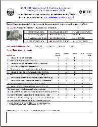
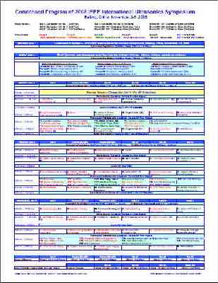
Condensed Technical Program and Some Statistics on Technical Program, Attendence, Registration, Short Course, and Proceedings:
|
Condensed Program: |
Program Sheet: |
|
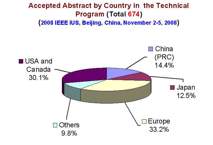
1. Abstract Accepted by Country: |
2. Conference Registration by Country: |
|
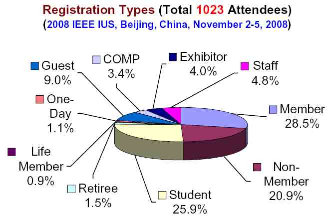
3. Conference Registration Types: |
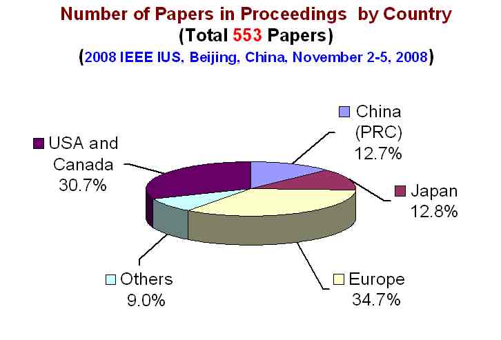
4. Proceedings Papers by Country: |
|
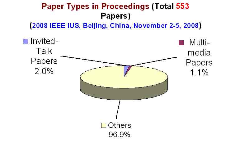
5. Paper Types in Proceedings: |
6. Abstract Statistics for Proceedings: |
|
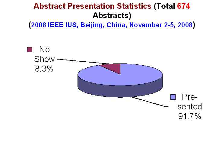
7. Abstract Presentation Status: |
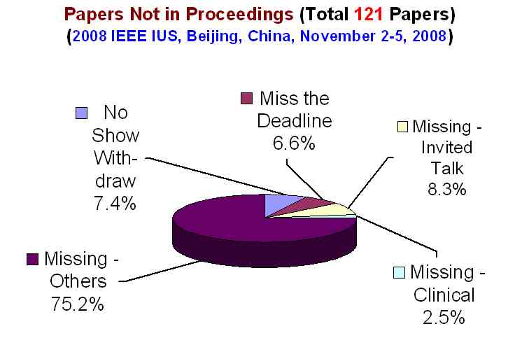
8. Papers Not in Proceedings: |
|
9. Short Course Registration Types: |
10. Short Course Registration by Courses: |
Home Contact the webmaster, Dr. Jian-yu Lu, for questions. © Copyright 2006-2008 IEEE UFFC Society