

Home Click on Images above to Enlarge
Home
Click on Images above to Enlarge
2008 IEEE International Ultrasonics Symposium (IUS)
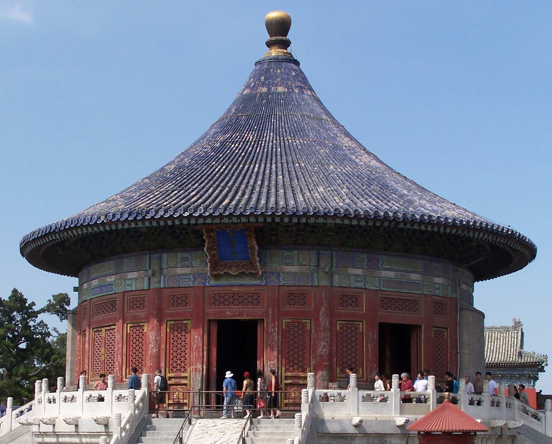
Beijing International Convention Center (BICC)
Beijing, China, November 2-5, 2008 (View: Conf. Photos/Videos and ![]() Beijing Photos
Beijing Photos ![]() )
)
|
|
Sponsored by the IEEE Ultrasonics, Ferroelectrics, and Frequency Control Society |
|
Student Paper Competition Finalists (21 in Total)
Student Paper Competition Finalists:
21 Student Paper Competition finalists have been selected during the 2nd Technical Program Committee meeting that was held from June 14-15, 2008 in Chicago, Illinois, USA. The finalists should check the award selection criteria and the requirements for their presentations at the link: Student Paper Competition.
Photos and videos of the Student Paper Competition Finalists and the award winners are now available via "Conference Photos/Videos".
Note: The deadline, Friday, August 1, 2008, to resolve with your employers or any other concerned body for any intellectual property rights of the researches in the abstracts (please see "Abstract Results" for detail) has passed. The full abstracts of all the Student Paper Competition finalists are now available through the links below.
Finalist #1.1 (PS001-01) (3F-2):
Title: Design of Catheter for Combined Intravascular Photoacoustic and Ultrasound Imaging
Bo Wang (Presenter), Andrei Karpiouk, and Stanislav Emelianov, Biomedical Engineering, University of Texas at Austin, Austin, TX, USA. (Abstract ID: 309)
Abstract:
Background, Motivation and Objective:
Intravascular photoacoustic (IVPA) imaging is a promising imaging tool for detecting and differentiating the atherosclerotic plaques. Previously, we have demonstrated the utility of intravascular photoacoustic imaging using a laboratory system where the excised arterial tissue sample was irradiated with the laser beam from the outside while the intravascular ultrasound (IVUS) imaging catheter, inserted into the lumen, was used to receive the photoacoustic signal. However, for in-vivo IVUS and IVPA imaging, a combined catheter consisting of IVUS transducer and light delivery system is needed. In this paper we report our initial experience towards design and fabrication of a catheter capable of simultaneous IVPA and IVUS imaging.
Statement of Contribution/Methods:
The combined IVUS/IVPA imaging catheter was built based on a clinical, 40 MHz, single element IVUS catheter (Boston Scientific, Inc.). A 0.6 mm diameter optical fiber was used for light delivery. The proximal end of the fiber was coupled with a laser system. The distal tip of the optical fiber was polished at a 45 degree angle and placed inside a quartz tube. Both ends of the tube were sealed with epoxy to retain air around the fiber tip. As a result, such fiber assembly, when submersed into water, was irradiating the light sideway. The IVUS catheter was then attached to the optical fiber such that the ultrasound beam from the transducer and the laser beam from the optical fiber were aligned. To test the combined IVUS/IVPA imaging catheter, a model of the atherosclerotic vessel was fabricated. Specifically, within the otherwise homogeneous polyvinyl alcohol background, three 0.4% graphite inclusions of 1 mm diameter were positioned at different depths inside of the approximately 6-mm thick vessel wall to simulate various plaques in the artery. During imaging studies, the catheter was inserted into the lumen, and the phantom was rotated using a stepper motor. At each angular position, both photoacoustic and ultrasound A-lines were collected by a 14-bit GAGE A/D card operating at 200 MHz sampling rate. The IVUS and IVPA images were formed off-line from 256 equally spaced beams.
Results:
The IVPA images of the phantom obtained using the combined IVUS/IVPA catheter clearly identifies the inclusions located at specific depths. At the same time, co-registered IVUS images visualized the structure of the phantom. Finally, combined IVUS/IVPA images further outlined the location and extent of the inclusions within the vessel wall.
Discussion and Conclusions:
Overall, IVPA and IVUS images of sufficient quality were obtained using the initial prototype of the combined IVUS/IVPA catheter. Using optical fibers of smaller diameter, the size of the combined catheter can be further reduced. Therefore, our studies suggest that optical fibers can be used to deliver enough optical fluence for intravascular photoacoustic imaging of the vessel. Furthermore, other approaches in design of IVUS/IVPA imaging catheter will be discussed.
Finalist #1.2 (PS002-02) (2F-6):
Title: Intra-Vascular Ultrasound (IVUS) Delivery of DNA Via Microbubble Carriers to an Injured Artery In vivo
*Linsey C. Phillips (Presenter), *Alexander L. Klibanov, **Doug K. Bowles, *Brian. R. Wamhoff, and *John A. Hossack, *University of Virginia, Charlottesville, VA, USA, **University of Missouri, Columbia, MO, USA. (Abstract ID: 1094)
Abstract:
Background, Motivation and Objective:
The most common therapy for narrowed, atherosclerotic arteries is balloon angioplasty and is often followed by stent placement. This procedure causes injury to the vessel wall and over time, cells from the artery wall (primarily smooth muscle cells) proliferate in response to injury and re-occlude the vessel (restenosis). A novel therapy to prevent restenosis involves the use of delivering an anti-proliferative gene via microbubbles which are ruptured via catheter-based intravascular ultrasound at the site of vessel injury. Insonation of microbubbles by ultrasound has been shown to increase gene delivery at low frequencies. We hypothesize that plasmid DNA encoding a reporter gene (red fluorescent protein, RFP) can be delivered to a pig carotid artery wall in vivo using cationic microbubble carriers and intravascular ultrasound.
Statement of Contribution/Methods:
Cationic microbubbles were formed during ultrasonic dispersion of decafluorobutane gas in an aqueous micellar mixture of phosphatidylcholine, PEG stearate, and distearyl trimethylammonium propane. Negatively charged DNA plasmids expressing red fluorescent protein (CMV-RFP) were electrostatically coupled to microbubbles.
A modified IVUS catheter (Boston Scientific) was positioned 1mm away from cells and translated over an area of 2cm2. In vitro application of 5 or 11 MHz Guassian pulses at a PNP of 120 kPa, and PRF of 5kHz was applied to cultured smooth muscle cells exposed to CMV-RFP plasmid bearing microbubbles for at total of 6 minutes.
Balloon angioplasty was performed on pig right carotid vessels (n=2) in vivo. (The pig is the gold standard for restenosis studies.) Following angioplasty, microbubbles were infused through a port hole in a catheter located 2cm upstream of the IVUS transducer. 5 MHz unipolar pulses (PRF = 5 KHz, PNP =120 kPa) were emitted from the IVUS catheter at the location of vascular injury (2cm in length) for a total of 4 minutes during plasmid conjugated-microbubble infusion. Three days following insonation, arteries were excised and processed for frozen sectioning and nuclei staining. Successful plasmid transfection was measured by fluorescent microscopy and quantified as % of vessel perimeter cells expressing RFP.
Results:
Ultrasound mediated gene delivery from microbubbles using IVUS in vitro resulted in 11.5 fluorescent cells/cm2 (<1%). (Previously reported ultrasound / bubble mediated gene delivery are typically low ~1-5%.) Cells exposed to plasmidmicrobubbles without US resulted in 0% transfection. Injured pig arteries exposed to microbubbles and ultrasound resulted in 23.3?6.0% transfection whereas contralateral controls resulted in 3.6?.6% transfection.
Discussion and Conclusions:
To the authors?knowledge, this is the first example of in vivo cationic gene delivery via IVUS, which has promise for localized intra-vascular gene therapy for preventing restenosis.
Finalist #1.3 (PS003-03) (3E-5):
Title: Quantitative Bladder Volume Assessment on the Basis of Nonlinear Wave Propagation
Egon J.W. Merks (Presenter), Nicolaas Bom, Nico de Jong, and Antonius F.W. van der Steen, Biomedical Engineering, Erasmus MC, Rotterdam, Netherlands. (Abstract ID: 291)
Abstract:
Background, Motivation and Objective:
Catheterization is the gold standard for bladder volume assessment, but it is invasive and introduces the risk of infections and trauma. To reduce the need for a urinary catheter, a new method has recently been introduced that non-invasively and instantaneously measures the volume of liquid filled cavities on the basis of nonlinear wave propagation with a single diverging acoustic beam. The method exploits the relatively higher nonlinear behavior of liquid compared to tissue.
Previously obtained results from using a fast-rotating phased array probe and high end echo system have proven the feasibility of the method. A 15 dB increase of the 2nd harmonic was observed on a 500 ml bladder phantom relative to a tissue-only phantom. In-vivo measurements on a bladder containing 450 ml urine showed an increase of 10 dB at the 2nd harmonic compared to an empty bladder. The objective of this study is to design a simple transducer that generates a single diverging acoustic beam and to obtain a calculation method that quantitatively relates the spectral contents of the received RF-data to the insonified liquid volume.
Statement of Contribution/Methods:
Progressive volume pulse-echo measurements were performed on healthy volunteers. The experimental setup included a custom multilayer transducer that is capable of generating sine wave bursts with centre frequency of 2 MHz and peak amplitudes of 500 kPa at the transducer surface, which could induce significant nonlinear wave propagation. The transducer bandwidth enabled receiving up to the 4th harmonic. To create the diverging acoustic beam, a defocusing lens was applied to the transducer. Volumes between 0 and 600 ml with 100 ml increments were measured. Reference measurements were performed with a commercial bladder volume instrument. Subject-specific acoustic loss and nonlinearity of the region anterior to the bladder influenced the volume estimation. A calculation scheme was applied that subtracted the nonlinear behavior of the anterior bladder region from the nonlinear behavior of the posterior bladder region, leaving only the fraction of nonlinearity (FON) contributed by the liquid region.
Results:
Linear regression analysis on the data obtained from the progressive volume measurements resulted in a slope of 4.6 L/FON and an intercept of 118 ml. The 95% confidence interval of the slope was [4.6 ?0.7]. By correcting for the nonlinear behavior of the anterior bladder region and looking at only the fraction of nonlinearity contributed by the liquid region, the relative standard deviation of the slope was reduced from 19.3% to 8.4% for the individual progressive measurements. Hence, the repeatability of the method increased significantly.
Discussion and Conclusions:
A calculation method was developed that quantitatively relates the spectral contents of the received RF-data to the liquid volume present within a single diverging acoustic beam in-vivo.
This work was supported by the Dutch Technology Foundation (STW) under Grant 06652.
Finalist #1.4 (PS004-04) (2D-3):
Title: Microbubble dynamics in microvessels: Observations of microvessel dilation, invagination and rupture
Hong Chen (Presenter), Andrew A. Brayman, Michael R. Bailey, and Thomas J. Matula, Center for Industrial and Medical Ultrasound, Applied Physics Laboratory, University of Washington, Seattle, WA, USA. (Abstract ID: 609)
Abstract:
Background, Motivation and Objective:
The fundamental interaction of an acoustically activated microbubble with small blood vessels is poorly understood. Understanding this interaction is important for designing better imaging schemes, and for targeting and drug delivery applications. High speed microscopy provides a tool to study interaction and response mechanisms.
Statement of Contribution/Methods:
Following an approved U.W. IACUC protocol, ultrasound contrast agent microbubbles, Evan’s blue and fluorescent dyes were perfused into the rat mesentery. These tissue samples were harvested for ex vivo observation. Evan’s blue was used to facilitate identification of microvessels and also as an indicator of blood vessel permeability changes. Fluorescence images were taken to examine the integrity of blood vessels. Tissue samples were exposed to short pulses of 1 MHz ultrasound. 14 high-speed microphotographic images were acquired for each experiment with shutter speeds of 50 ns and each image separated by 150 ns.
Results:
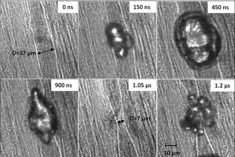
At low acoustic negative pressure (~1.5 MPa), bubble expansion caused microvessel dilation by approximately 1.2x. During bubble collapse, the vessel invaginated to approximately 0.9x of its original diameter (11 ?m). At high negative pressure (near 11 MPa), the vessel dilated by approximately 2.5x, followed by invagination of 0.4x of its original diameter (17 ?m). Vessel dilation and invagination were correlated temporally with bubble growth and collapse. At high pressure, the bubble and/or its fragments could be observed outside the original vessel, suggesting that the vessel had ruptured at some point. Vessel damage was also inferred by observation of fluorescent dye extravasation. An example of vessel dilation, invagination, and rupture can be seen in the following figure (pixel intensity values in the region around the blood vessel wall have been enhanced).
Discussion and Conclusions:
Our observations confirm some aspects of previous modeling and observational findings. However, direct observation of ultrasound-induced vessel invagination appears novel, and may be an important mechanism related to vessel damage. It remains uncertain if the vessel was damaged during dilation, invagination, or from a violent bubble collapse. It’s possible that both dilation and invagination contribute to vascular rupture. Work supported by NIH (5R01EB000350 and P01DK43881).
|
|
Finalist #1.5 (PS005-05) (2H-4):
Title: Non-invasive thrombolysis induced by histotripsy pulsed cavitation ultrasound therapy
*Adam Maxwell (Presenter), *Charles Cain, **Hitinder Gurm, ***J. Brian Fowlkes, and *Zhen Xu, *1Department of Biomedical Engineering, University of Michigan, Ann Arbor, Michigan, USA, **Department of Internal Medicine, University of Michigan, Ann Arbor, Michigan, USA, ***Department of Radiology, University of Michigan, Ann Arbor, Michigan, USA. (Abstract ID: 68)
Abstract:
Background, Motivation and Objective:
Blood clot formation is an essential response to injury but can be the cause of many cardiovascular diseases. Current treatments to remove blood clots (thrombolysis) include thrombolytic drugs and/or catheter-based techniques, both of which have significant drawbacks including risks of excessive bleeding and infection. Our goal is to develop a noninvasive thrombolysis method based on Histotripsy, a technique that mechanically fractionates soft tissue using controlled ultrasound cavitation. This paper investigates the feasibility and efficacy of this new approach to thrombolysis.
Statement of Contribution/Methods:
Blood clots were formed in-vitro from whole porcine blood by adding CaCl2 solution. Clots were placed in a 6 mm diameter LDPE tube and treated by histotripsy. The treatment targeting and monitoring were guided by ultrasound imaging. The histotripsy treatment consisted of 5 cycle ultrasound pulses delivered at a 1 kHz pulse repetition frequency and peak negative pressures of up to 14 MPa. Clots were treated until completely dissolved. Acoustic backscatter during treatment was collected for cavitation detection. Clots were also treated under flow rates up to 50 cm/sec in a circulatory model. To evaluate possible vascular damage, clots were treated in excised canine aorta and vena cava and histology of the vessels was examined for damage.
Results:
Histotripsy can completely fractionate a clot weighing 300 mg (4 mm in diameter and 2 cm in length) in ~0.5 ?5 minutes (mean = 2.7 minutes, n = 32 clots). Histotripsy thrombolysis was initiated at peak negative pressures ? 8 MPa, and only after initiation of a cavitating bubble cloud was detected. The thrombolysis rate (clot weight/treatment time) increased with increasing pressure. Histotripsy fragmented the clot into debris no larger than 60 ?m in diameter, with over 90% (by volume) of the debris having diameters < 8 ?m. The treated vessels were intact upon initial histological evaluation. Histotripsy thrombolysis was effective both in high flow and static environments. The treatment targeting and progress can be clearly seen on an ultrasound image. Moreover, we observed that clot fragments are attracted to the vicinity of the bubble cloud, and can be trapped and further fragmented at the focus.
Discussion and Conclusions:
Our results suggest that histotripsy is an effective and efficient non-invasive method for thrombolysis guided by real-time imaging. Most clot debris fragments generated are smaller than red blood cells. Large clot fragments can be trapped near the bubble cloud and further fractionated. This phenomenon is possibly due to a particular fluid flow pattern created by cavitation-induced microstreaming. We plan to use this property to create a Non-invasive Embolization Trap (NET) to prevent embolization caused by escaping clot fragments. These results suggest that histotripsy has the potential to emerge as a safe and effective non–invasive thrombolytic.
Finalist #1.6 (PS006-06) (2I-5):
Title: Reaching the optimal focusing and steering capabilities of transcranial HIFU arrays based on time reversal of acoustically induced cavitation bubble signature
*Jerome GATEAU (Presenter), **Laurent MARSAC, *Mathieu PERNOT, *Jean-Francois AUBRY, *Mickael TANTER, *Mathias FINK, *Laboratoire ondes et Acoustique, INSERM, CNRS UMR 7587, ESPCI, PARIS, France, **SUPERSONIC IMAGINE, Aix-en-Provence, France. (Abstract ID: 248)
Abstract:
Background, Motivation and Objective:
Brain treatment with High Intensity Focused Ultrasound (HIFU) can be achieved through the skull by multichannel arrays using time-reversal focusing. Such a method requires a reference signal either sent by a real source embedded in brain tissues or computed from a virtual source, using CT based simulations. This non-invasive computational method allows precise focusing, but discretization and modeling errors can result in a reduction of the accessible acoustic pressure at focus in comparison with real experimental time-reversal using an implanted hydrophone. The goal of this study is to demonstrate the feasibility of reaching the optimal focusing based on the initial corrections obtained from CT-scan simulations. The optimal acoustic pressure at focus is recovered by inducing a cavitation bubble through the skull bone and using its ultrasonic emission for time-reversal transcranial focusing. The potential of this technique for improving both transcranial focusing and electronic beamsteering performances is investigated.
Statement of Contribution/Methods:
Ex vivo experiments are performed on a half skull immersed in a degassed water tank maintained at 37 C. The ultrasound array is composed of 136 high-power individual transducers (central frequency 1MHz) mounted on a spherical surface with a semi random distribution. The simulation uses a 3D finite differences code and a model of the half skull based on CT data. Cavitation events occur in an agar gel, phantom for in vivo bubble formation. The pressure field at 1MHz is scanned at low amplitude levels with a hydrophone mounted on a 3D gantry.
Results:
Ex vivo CT guided simulations allowed us to reach, at the geometrical focus of the array, 83% of the optimal pressure (hydrophone based time reversal). Cavitation bubbles were then created transcranially at this location with computed emission pulses. The 1MHz component of a single bubble acoustic emission was selected, time reversed and reemitted, restoring a mean pressure ratio of 96% (+/- 2%). The new focal peak, i.e the location of the cavitation event, was localized in the -2dB focal area of the initial pulse corresponding to a 0.5 mm uncertainty. When performing electronic steering from a reference signal optimally focusing at the geometrical focal point, 90% of the optimal pressure is still reached up to 8 mm away to the initial position in the focal plane. With six reference signals from cavitation bubble spots equally distributed on a 6 mm radius circle, this area was extended to 12 mm. Such cavitation bubbles were generated using electronic steering.
Discussion and Conclusions:
A new non-invasive method to correct skull aberrations has been validated. From CT images based simulations, the focusing was restored through the skull by inducing a cavitation bubble at the targeted location, and the corrected zone was extended by electronic beam steering and discreet bubbles generation This method should greatly benefit transcranial brain therapy.
Finalist #1.7 (PS007-07) (2K-5):
Title: High Frame Rate Adaptive Imaging Using Coherence Factor Weighting and the MVDR Method
Shun-Li Wang (Presenter) and Pai-Chi Li, National Taiwan University, Taipei, Taiwan. (Abstract ID: 1083)
Abstract:
Background, Motivation and Objective:
Adaptive imaging has been extensively studied. Although some success has been demonstrated, these approaches generally are not suitable for high frame rate (HFR) imaging where broad transmit beams are required. In this study, we propose an effective adaptive imaging method suitable for HFR imaging based on coherence factor (CF) weighting and the minimum variance distortionless response (MVDR) method.
Statement of Contribution/Methods:
The CF is a focusing quality index estimated from receive-channel data. It is the ratio between the energy of the coherent sum to the total incoherent energy. This method is an adaptive weighting technique in which the amplitude of each image pixel is weighted by the corresponding CF such that the unwanted sidelobes are reduced. Direct implementation of the CF weighting in HFR imaging does not provide satisfactory results because broad transmit beams required for HFR imaging affect accuracy of CF calculations. In this study, we solve this problem by applying the MVDR method to the delayed channel data. Specifically, the MVDR method is used for angle of arrival estimation. The beam sum data are then weighted by the estimated CF.
Results:
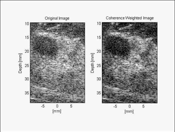
A synthetic transmit aperture method is used for HFR imaging. Only 8 firings are required to form an image. Both simulations and clinical breast imaging data are used. In the simulations, an anechoic cyst phantom is imaged with a maximum ?/2 near field phase screen. The correlation-based method proposed by Flax/O?Donnell is also implemented for performance benchmarking. The contrast and the contrast-to-noise ratio (CNR) improvements are 7.7 dB and 39.2% with the proposed method, respectively. Only 2.1 dB and 21.4% improvements are achieved using the correlation-based method. Clinical breast data are also acquired using a programmable array system. The following figures show images of a fibroadenoma lesion (left: original image, right: with the proposed method). With the proposed data, the contrast enhancement is 3.3 dB and the CNR enhancement is 13.4%.
Discussion and Conclusions:
The proposed method combines CF weighting with the MVDR method. Simulations and clinical breast data are used to demonstrate the image quality improvement. Even for HFR imaging with only 8 firings per image, effective contrast enhancement and better lesion boundary can be achieved. Efficacy of the proposed method is clearly demonstrated.
|
|
Finalist #1.8 (PS008-08) (1K-5):
Title: Estimation of Valvular Regurgitation Area by 3D HPRF Doppler
*Torbjørn Hergum (Presenter), *Thomas Renhult Skaug, **Knut Matre, and Hans Torp, *Department of circulation and medical imaging, Norwegian University of Science and Technology, Trondheim, Norway, **Institute of Medicine, University of Bergen, Bergen, Norway. (Abstract ID: 1040)
Abstract:
Background, Motivation and Objective:
Determining the severity of leakage through a heart valve is important, but difficult. Two of the parameters which are clinically interesting in this regard are the area and the geometry of the lesion. Current practice for non-invasive measurement of the severity of valvular regurgitation is qualitative, and based upon using color flow- and spectral Doppler techniques.
Statement of Contribution/Methods:
In search for quantitative measurements of regurgitant severity we used 3D high pulse repetition frequency (HPRF) color flow imaging to measure the Doppler signal from multiple beams distributed over the laminar vena contracta region near the orifice. A steep clutter filter was used to separate the jet flow Doppler signals from the Doppler signals of the slowlymoving blood of the ambiguous sample volumes.
The power from the closely spaced ultrasound beams are summed to yield the total Doppler power, which is known to be proportional to the amount of blood moving above the clutter-filter cutoff velocity. The cross sectional area of the jet was found by scaling the summed Doppler power from these beams using both a-priori knowledge of the lateral extent of the beams and a reference beam which is completely covered by the orifice.
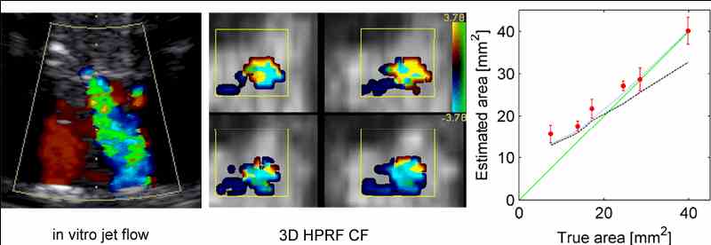
Both in vitro trials and computer simulation have been used for validation. The in vitro measurements were made using a pulsatile flow phantom holding porcine valves with six different holes, ranging from mild to severe mitral regurgitation. The method can be applied to other high-velocity valvular jets.
Results:
The mean value and standard deviation from the in vitro trials are plotted as red in the figure showing true area vs. estimated area. Two computer simulations are also included in the figure, the dashed and dotted lines are simulations respectively with- (blue) and without (black) stochastic variation.
Discussion and Conclusions:
Small holes of sizes comparable to a single ultrasound beam are overestimated as expected from simulations, and the estimates of the larger holes fits well with the line of identity (green). According to the stochastic simulations the method should underestimate the area of large orifices, but this is not seen in the in vitro data. Regardless of this the in vitro data enables us to distinguish between the different regurgitation degrees.
|
|
Finalist #1.9 (PS009-09) (2E-6):
Title: Image-Guided Refocusing of Dual-Mode Ultrasound Arrays(DMUAs)
John Ballard (Presenter) and Emad Ebbini, University of Minnesota, USA. (Abstract ID: 384)
Abstract:
Background, Motivation and Objective:
A major advantage of imaging with dual-mode ultrasound arrays (DMUAs) is their inherent registration between imaging and therapeutic modes during image-guided surgery which allows for image-based feedback for refocusing the therapeutic beam. Specifically, this capability is critical in image-guided thoracic surgeries where the target is partially obstructed by the rib cage, thus limiting the access and distorting the geometrically-focused high-intensity focused ultrasound (HIFU) therapeutic beam.
Statement of Contribution/Methods:
Images obtained with single-transmit focus (STF) imaging, in which the therapeutic beam is used at diagnostic levels, allow the user to select target and critical locations for optimizing the power deposition. We have developed an optimal refocusing method that takes advantage of the acoustic window of the intercostals spacing in order to minimize the power deposition over the critical regions (ribs) while maintaining or improving the power deposition at the target location
(tumor).
Results:
The algorithm is verified experimentally with a 64-element 1MHz DMUA, in an attenuating tissue mimicking phantom (~.5 dB/cm/MHz) with embedded Plexiglas ribs. Thermocouples are used to measure sub-therapeutic temperatures across the ribs and at the target location before, during and after 5 seconds of HIFU exposure for both the geometric focusing and the optimized refocusing while normalizing the driving power for both cases. An increase of normalized temperature (per watt of input power) greater than 20% was observed at the target after refocusing. At the same time, a reduction in normalized temperature rise across the ribs was greater than 60%. Statistics showed that the maximum variance between measurements when the experiment was rerun a minimum of 5 times for each case was approximately 5%. In addition, STF images taken with the refocused HIFU beam showed increased echogenicity at the target and reduced echogenicity at the ribs. This can be quantified by the intensity of the grayscale images. These images show a typical improvement of 5 dB at the focus with a reduction of 2dB across the ribs.
Discussion and Conclusions:
The results show that STF DMUA imaging provides suitable feedback for refocusing the HIFU beam in the presence of strongly scattering targets. The robustness and repeatability of the algorithm were demonstrated by embedding the ribs within a block tissue-mimicking phantom to approximate realistic conditions and repeating each experiment multiple times. The results also show that grayscale STF images themselves provide useful feedback on the improvement in the quality of the refocusing beam, i.e. the relative echogenicity of the ribs is reduced upon refocusing indicating reduction in incident power. These results are generally consistent with the directly measured temperatures at the target and rib locations.
Finalist #2.1 (PS010-10) (5C-2):
Title: The Detection of Chemical and Biological Analytes Using a Monolithic Spiral Coil Acoustic Transduction Sensor
Donald McCann (Presenter), Mitchell Wark, Paul Millard, David Neivandt, and John Vetelino, University of Maine, Orono, ME, USA. (Abstract ID: 131)
Abstract:
Background, Motivation and Objective:
The monolithic spiral coil acoustic transduction (MSCAT) sensor platform is a novel bulk acoustic wave (BAW) device which is excited by a gold spiral coil antenna photolithographically deposited on one side of an AT-quartz wafer. The MSCAT platform can operate at very high frequencies by efficiently exciting high harmonic transverse shear modes with the application of a high frequency RF signal to the spiral coil. Since one surface of the MSCAT device is bare, this device can be used as a sensing platform upon which one deposits analyte selective chemical or biological films. The bare surface allows the detection of analyte induced mechanical (mass and viscoelasticity) and electrical (conductivity and dielectric constant) property changes in the film. In order to demonstrate the applicability of a MSCAT device as a sensor, the MSCAT platform is coated with biological and chemical films selective to Escherichia coli (E. coli) O157:H7 (hereafter referred to as E. coli), the E. coli strain most often responsible for serious illnesses in humans, and saxitoxin (STX), the most dangerous neurotoxin associated with shellfish poisoning stemming from red tide, respectively.
Statement of Contribution/Methods:
A method for optimizing the number of turns and coil width and spacing of the MSCAT's antenna was developed using the Box-Behnken design method. MSCAT sensing platforms were then coated with biological films selective to E. coli based on antibody-antigen interactions and chemical films selective to STX based on the 18-crown-6 ether. Each MSCAT sensor was the exposed to E. coli and STX and the changes in resonant frequency were monitored.
Results:
It was found that the most critical parameter in achieving efficient operation of the MSCAT device was the coil width. The MSCAT sensor operating at its fundamental frequency (5 MHz) was exposed to E. coli and exhibited a frequency shift approximately five times greater than similar tests performed with quartz crystal microbalance (QCM) sensors. In order to determine the lower detection limit and resolution of the MSCAT sensor, the sensor was operated at its 11th harmonic (55 MHz) and exposed to decreasing concentrations of E. coli. The resonant frequency was then monitored to obtain a dose response curve. The MSCAT sensor was able to detect E. coli in concentrations as low as 104 microbes/mL, 2 orders of magnitude lower than the QCM sensor. Similar results relating to the detection limit and resolution were also obtained for STX.
Discussion and Conclusions:
A method for optimizing the MSCAT sensors' spiral coil antenna geometry was performed and it was shown that the coil width was the most critical parameter. The MSCAT was found to be significantly more sensitive than the QCM sensor due to the fact that it can detect both electrical and mechanical property changes and operate at high frequencies. Since the MSCAT has been excited up to the 81st harmonic, the MSCAT device may also be used in high frequency resonator applications.
Finalist #2.2 (PS011-11) (5G-5):
Title: Improving the Bandwidth of Air Coupled Capacitive Ultrasonic Transducers Using Selective Networks
*Sean Mc Sweeney (Presenter) and **WMD Wright, *Electrical and Electronic Engineering Dept, University College Cork, National University of Ireland, Mallow, Cork, Ireland, **Electrical and Electronic Engineering, University College Cork, National University of Ireland, Cork, Cork, Ireland. (Abstract ID: 589)
Abstract:
Background, Motivation and Objective:
One of the key limitations on using CUT (Capacitive Ultrasonic Transducers) and cMUTs[1] (Capacitive Micromachined Ultrasonic Transducers) in air is their relatively narrow bandwidth which although superior to that of current piezoceramic devices[2] could be improved. Most air coupled capacitive devices could benefit hugely through the use of selective networks[3] for bandwidth expansion, resonance reinforcing, or a combination of both. This work has investigated the application of pole/zero manipulation techniques to modify and enhance the transmission characteristics of capacitive transducers through front end mounted components. The main objective was to positively enhance the performance characteristics of capacitive transducers.
Statement of Contribution/Methods:
A modified electrical equivalent circuit for CUTs to include the selective networks used was developed. The work assessed the effects of a tuned amplifier on the passband of the devices studied and then focused on more complicated network designs for enhancement. Simulations of the effects of the networks on the devices using equivalent circuit models were carried out and the response curves to pulsed operation were calculated and compared to experimental measurements from a pair of fixed CUTs with a combined centre frequency of 280kHz and 3dB bandwidth of 160kHz.
Results:
Increases in centre frequency of 25% and 3dB bandwidth of 77% using a single tuned amplifier were obtained. Resonance reinforcing, resonance shifting and ripple suppression were also studied through the manipulation of the q factor and pole location of such an amplifier. Performance enhancements were studied for a number of CUT aperture sizes and membrane thicknesses and a comparative study of the theoretical and experimental effect of these variations was conducted. With the appropriately designed network, enhancement of peak resonance with a simultaneous bandwidth expansion was obtained at the expense of other operating parameters such as stopband ripple. Simulations of more complicated circuit designs using equivalent circuit models of capacitive devices[4,5] showed that the maximum level of passband ripple observed for the bandwidth expansion method using a single tuned amplifier was reduced while achieving simultaneously the same 3dB results.
Discussion and Conclusions:
The implication for bandwidth expansion of a capacitive transducer through selective network design is significant, allowing increased resolution in imaging systems, ultrasonic ranging and non destructive evaluation. Significant improvements have been observed without additional signal manipulation, through digital means or otherwise, in certain transmission properties of the devices. Future work will expand on the enhancement of capacitive transducers through the use of hybrid resonator circuits and other related methods.
Finalist #2.3 (PS012-12):
Title: Dynamic focusing thorough arbitrary geometric interfaces
Montserrat Parrilla (Presenter), Jose Brizuela, Jorge Camacho, Alberto Ibañez, Patricia Nevado, and Carlos Fritsch, Instituto de Automática Industrial (CSIC), La Poveda (Arganda), Madrid, Spain. (Abstract ID: 154)
Abstract:
Background, Motivation and Objective:
The use of array technology in Non-Destructive-Testing (NDT) applications requires setting the focal laws in emission and in reception for every array element. Frequently wedges are inserted or the inspection of the part is carried out by immersion. In these cases, the presence of interfaces complicates the focal law computing task due to refraction effects. If the number of foci is limited (i.e., single focus in emission and in reception), the Fermat’s principle can be applied to accurately compute the focal laws.
However, state-of-the art equipment allows dynamic focusing, which provides increased resolution, signal to noise ratio and contrast. With dynamic focusing, the number of focal laws to be computed increases by two or three orders of magnitude and the computing time by the Fermat’s principle increases accordingly.
The main objective of this work is to present new methods which provide the dynamic focusing focal laws with the required accuracy and less computing burden.
Statement of Contribution/Methods:
Several methods of focal law computing thorough arbitrary geometry interfaces are analyzed. As a background, the general purpose method based on the Fermat principle is described. Then, a new approach, the Fast Focal Law Computing (FFLC) technique is presented. It is based in solving an equation by an iterative method. It is shown that, for most applications and following the proposed processing method, no iterations are required, being nearly as fast as a closed formula method.
The Fermat and the FFLC methods are compared with regard to accuracy and computing time for dynamic focusing of a diversity of interface shapes. Then, both methods are applied to compute the dynamic focal laws into aluminum parts with artificial flaws, using a 5 MHz transducer. Wedges are applied for planar interfaces and water immersion is used for shaped parts with an irregular geometry.
Results:
It is shown that the proposed FFLC algorithm obtains the dynamic focal laws in about 1/20 the computing time required by the conventional Fermat’s principle application. This represents getting the equipment ready for inspection in a few seconds, instead of several minutes. Furthermore, focusing errors of the FFLC are very small and comparable to those produced by the standard method. Thus, image quality is not impaired in spite of the low time spent in computing the focal laws.
Discussion and Conclusions:
The FFLC is presented and analyzed. It provides a fast method to compute focal laws for dynamic focusing into parts of arbitrary shape. Images obtained with the computed focal laws are of the same quality that those obtained with more costly methods. This is due to the very low timing errors produced by the FFLC technique.
Finalist #3.1 (PS013-13) (6E-5):
Title: Wireless Drive of a Piezoelectric Plate by Dipole Antenna
Satyanarayan Bhuyan (Presenter) and Junhui Hu, Nanyang Technological University, Singapore. (Abstract ID: 72)
Abstract:
Background, Motivation and Objective:
In most applications of piezoelectric devices, electric energy is applied to the devices via lead wires soldered on the electrodes of piezoelectric components. But the lead wires may fall off at large vibration and high input voltage, and this causes the breakdown of piezoelectric devices. Thus, there is a need to introduce a wireless approach to apply electric energy to the piezoelectric devices. Wireless drive of a piezoelectric plate using an electric dipole antenna is explored in this work.
Statement of Contribution/Methods:
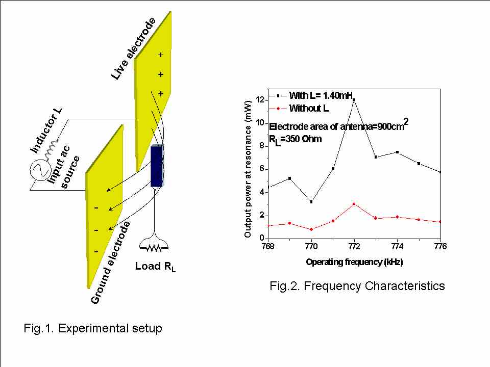
To transmit relatively large electric energy to a piezoelectric plate, an electric dipole antenna in series with an inductor is used as shown in Fig. 1. The ac electric field, produced by plate-shaped live and ground electrodes of the antenna is transmitted to the piezoelectric plate placed 6 mm away form antenna plane. The separation between antenna electrodes is 5 cm. The electric resonance of dipole antenna with an inductor generates a large voltage across the dipole antenna.
Results:
Fig. 2 shows the frequency characteristic of the output power of the piezoelectric plate operating in the thickness mode. At resonance frequency 772 kHz of the plate, a maximum output power of 12mW is achieved when the dipole antenna is in series electric resonance with an inductor because of the large voltage 1436Vrms across the antenna for an input voltage source of 150Vrms. The power transmitted to the load drops as the plate is detuned from resonance. An equivalent circuit of the wirelessly driven piezoelectric plate operating in the thickness mode has been developed. It is known that the circuit has a current source, resulting from the electric field which is different from the conventional piezoelectric plate driven by a voltage applied via lead wires.
Discussion and Conclusions:
A piezoelectric plate operating in the thickness mode is wirelessly driven by the electric field generated by a dipole antenna. At resonance a maximum output power of 12mW is achieved with an electrode area of 900 cm 2, input source voltage of 150Vrms, and 6mm from the antenna plane. An equivalent circuit of the wirelessly driven piezoelectric plate is derived which has a current source, resulting from the external electric field.
|
|
Finalist #3.2 (PS014-14) (6H-5):
Title: Towards thin film complete characterization using picosecond ultrasonics
Pierre-Adrien Mante (Presenter), Arnaud Devos, and Jean-François Robillard, IEMN-CNRS, France. (Abstract ID: 593)
Abstract:
Background, Motivation and Objective:
Mechanical characterization of thin films is a main issue in the microelectronic industry. The knowledge of these properties is necessary in many fields such as copper line interconnection and bulk acoustic wave resonators. A few techniques are reliable at this scale. Nano indentation or conventional laser-ultrasonic techniques can’t be effective in film thinner than 500 nm. Picosecond ultrasonics can also be used for thin film characterization. It is an efficient method to excite and detect vibrations within a thin film. A strong optical pulse warms a material surface, which leads to the creation of an acoustic wave propagating at the sound velocity. The waves propagation is longitudinal and it modifies the optical properties of the material. These modifications can be detected by a second time-shifted optical pulse.
Statement of Contribution/Methods:
In this technique we use a metallic very thin film as a transducer and only longitudinal waves can be generated. Due to that the full mechanical properties of thin layer cannot be measured. Here we show that thanks to a nanostructuration of the transducer, in-plane propagating waves are added using the same experimental setup. In the case of an isotropic medium, we have now access to all the acoustic properties.
Results:
We realized and studied 2D lattices of metallic nanocubes using e-beam lithography deposited onto the thin film to be charaterized. In a first experiment we will present results obtained on a 600nm-thick silica film.
Discussion and Conclusions:
Experiments were performed both on the lattices and out of the array of nanocubes. We respectively obtained the Rayleigh's velocity and the longitudinal velocity of silica. Then we can deduce Poisson's ratio and Young's modulus of silica: E=72GPa and nu=0.16, which is in very good agreement with literature. This first result demonstrates that we are able to extract longitudinal sound velocity, Rayleigh’s velocity, Young's modulus and Poisson's ratio in submicronic layers. Further results obtained on other materials isotropic and anisotropic will be also presented.
|
|
Finalist #3.3 (PS015-15) (6H-6):
Title: Simultaneous observation of induced longitudinal and shear acoustic phonons by Brillouin Scattering
*Yasuhiro Yoshida (Presenter), *Mami Matsukawa, and **Takahiko Yanagitani, *Faculty of Engineering, Doshisha University, Kyotanabe, Japan, **Department of Applied Physics, Nagoya Institute of Technology, Nagoya, Japan. (Abstract ID: 1015)
Abstract:
Background, Motivation and Objective:
Brillouin scattering measurement is a nondestructive method for measuring acoustic wave velocity at minute part of the material. This technique also enables us to measure longitudinal and shear wave velocities simultaneously. However, the measurement accuracy of the velocities is lower than those of other method such as pulse-echo measurement. This is mainly due to the weak Brillouin light scattering from the thermal phonons. In this study, we propose the use of induced longitudinal and shear waves for solving this problem.
Statement of Contribution/Methods:
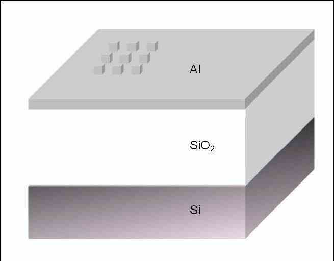
A c-axis tilted ZnO thin film transducer was deposited on side of the silica glass bar with the size of 3x10x35 mm3. Thus, excited continuous longitudinal and shear waves were propagated in the silica glass sample. Brillouin spectrum from silica glass sample were measured using RI?A scattering geometry [1].
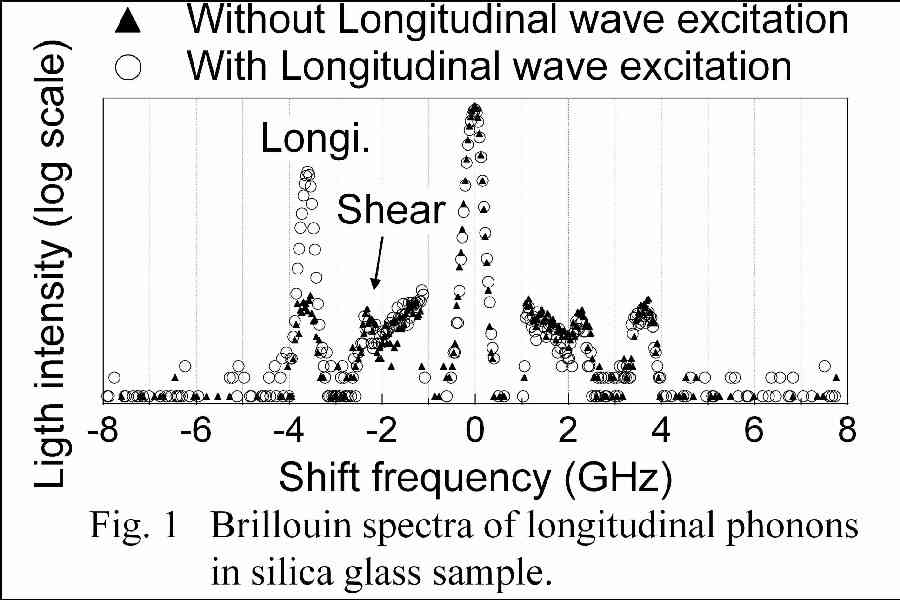
Results:
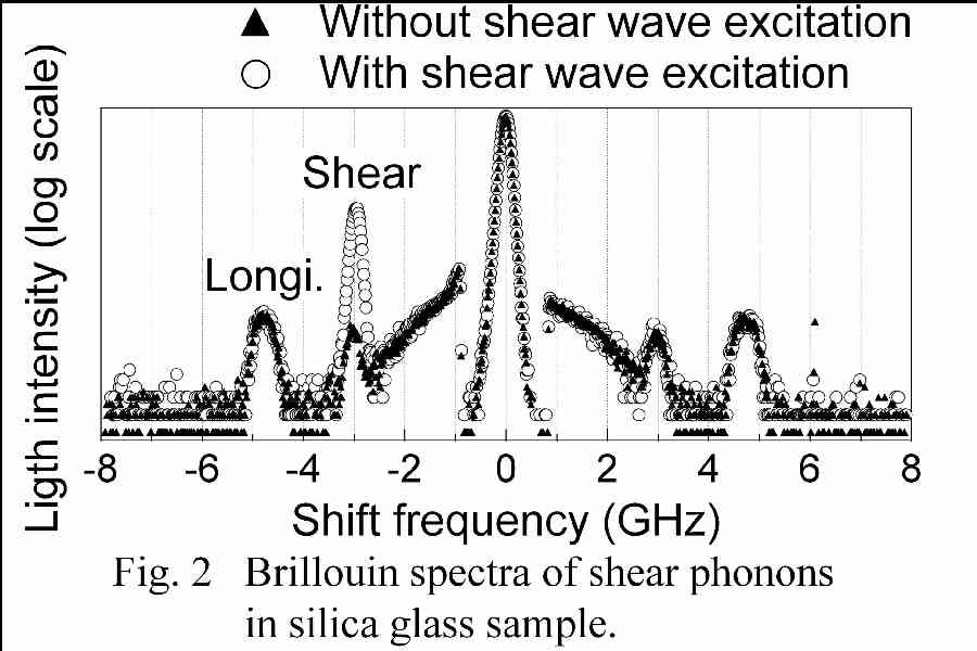
Figure 1 shows the Brillouin spectrum of longitudinal mode phonons observed without and with longitudinal and shear waves excitation. A pair of peaks observed at 3.6 GHz corresponds to the scattering from longitudinal mode phonons. This frequency is near the thickness extensional third overtone mode resonant of the film transducer. Strongly amplified Stokes peak is observed due to the excited longitudinal wave propagating in one direction. Also for shear mode phonons, amplified Stokes peak at 3 GHz (thickness shear fifth overtone mode resonant frequency) is observed as shown in Fig. 2.
Discussion and Conclusions:
This technique is useful for the sample which is easy to deteriorate because this technique realizes larger scattering even the use of lower laser power. Ref. [1]: J. K. Krüger et al., J. Phys. D: Appl. Phys 31 (1998) 1913.
|
|
|
Finalist #4.1 (PS016-16):
Title: Temperature Compensation of Thin AlN Film Resonators utilizing the Lowest order Symmetric Lamb mode
Gunilla Wingqvist (Presenter), Lilia Arapan, Ventsislav Yantchev, and Ilia Katardjiev, Solid State Electronics, Uppsala University, Uppsala, Sweden. (Abstract ID: 620)
Abstract:
Background, Motivation and Objective:
Micromachined Thin film plate acoustic wave resonators (FPAR) utilizing the lowest order symmetric Lamb wave (S0) propagating in highly textured 2?m thick Aluminum Nitride (AlN) membranes were successfully demonstrated [1]. The proposed devices had a SAW-based design and demonstrated Q factors of up to 3000 at a frequency of 900MHz as well as design flexibility with respect to the required motional resistance. A drawback of the proposed devices was the negative TCF of -20 ppm/K. Thus despite the promising features demonstrated, further device optimization is required.
Statement of Contribution/Methods:
In this work composite membrane employing the opposite temperature coefficients of delay of the DC sputtered AlN and the thermally grown SiO2 is used as a platform for the design of temperature compensated FPAR. The theoretical analysis, based on the Adler’s algorithm, revealed the possibility to achieve temperature compensation retaining the device electromechanical coupling. Further, the 1D equivalent model analysis suggested the use of Mo electrodes as a higher reflective alternative to the typically used Al in SAW-type reflecting gratings. Thus Mo being a material sustainable to electro-migration and with significantly smaller TCD, enables further device minimization as well.
Results:
The zero TCF devices demonstrated in here are synchronous type resonators fabricated on to composite AlN/SiO2 membrane consisting of relative thicknesses of d/?=0.166 and D/?~0.07, respectively. ?=12?m is the acoustic wavelength. The number of strips used in the reflectors is as small as 30 due to high reflectivity of the Mo electrodes. Q factors of around 1000 have been measured at a frequency of 850 MHz.
Discussion and Conclusions:
The latter is sufficient but slightly lower than the Q=1800 achieved for the synchronous noncompensated FPAR [1]. The observed reduction in Q is due to a slight non-uniformity of the thermally grown SiO2 layer caused by the limited selectivity of the Si/SiO2 Reactive Ion etching used for the membrane micromachning. Solutions of the problem are further suggested. In conclusion temperature compensated FPARs with reduced size and high Q are designed and micromachined on to low resisitve silicon wafers. Their potential applications include integrated frequency sorces as well as narrow band filters and gas sensors
Finalist #4.2 (PS017-17) (4J-2):
Title: A Full-Wave Analysis of Surface Acoustic Waves Propagating on a SiO2 Overlay/Metal Grating/Rotated YXLiNbO3 Substrate Structure
Yiliu Wang (Presenter), Ken-ya Hashimoto, Tatsuya Omori, and Masatsune Yamaguchi, Graduate School of Engineering, Chiba University, Chiba, Chiba, Japan. (Abstract ID: 217)
Abstract:
Background, Motivation and Objective:
The authors have recently reported a full wave analysis of piezoelectric boundary acoustic waves (PBAWs) propagating in a SiO2 overlay/Cu grating/rotated YX-LN substrate structure [1]. In the analysis, the finite element method is used for the grating region, while the spectral domain analysis is applied to an isotropic overlay region as well as a piezoelectric substrate region. The paper discusses in detail how the excitation and propagation characteristics of the shear-horizontal (SH) and Rayleigh-type PBAWs are dependent upon the Cu grating thickness, substrate rotation angle and metallization ratio.
The structure consisting of a SiO2 overlay of finite thickness is also directly applicable to the development of high performance SAW filters [2]-[3]. To the best of authors?knowledge, however, it seems that detailed discussions have not yet been made on the propagation characteristics of the SH- and Rayleigh-type SAWs.
Statement of Contribution/Methods:
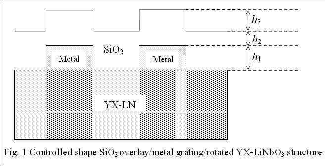
This paper describes a full wave analysis of the SH- and Rayleigh-type SAW propagation in a finite SiO2 overlay/metal grating/rotated YX-LN substrate structure shown in Fig. 1.
Results:
It is shown that the structure supports four types of propagation modes. Two modes concentrate their energy near the metal grating, and become the PBAWs when h2 gets infinite. Their electromechanical coupling is relatively strong even when h2 is large. One of the other two modes concentrates its energy near the top surface of the SiO2 layer. With an increase in h2, its propagation characteristics approach to those for the non-piezoelectric Rayleigh-type SAW on a semi-infinite SiO2 layer, rapidly losing its piezoelectric coupling. The remaining mode is the basis of a series of guided modes bounded in the SiO2 layer, in which the SiO2 layer behaves as a waveguide because of its low acoustic wave velocities.
Discussion and Conclusions:
Detailed discussions are made on the dependence of the propagation characteristics of these four modes on the design parameters such as the layer and grating thickness. It is also discussed how the propagation characteristics are affected by the SiO2 flatness denoted by h3 in Fig. 1.
|
|
Finalist #4.3 (PS018-18) (4I-5):
Title: Shear mode BAW resonator based on c-axis oriented AlN thin film
Evgeny Milyutin (Presenter), and Paul Muralt, Ecole Polytechnique Federale de Lausanne, Switzerland. (Abstract ID: 522)
Abstract:
Background, Motivation and Objective:
Thin film bulk acoustic wave resonators (TFBAR's) also showed potential as gravimetric sensors. In contrast to RF filters working with longitudinal modes, bio-medical applications usually require detection in a liquid, thus must employ shear modes. The principle has recently been successfully demonstrated with TFBAR devices employing tilted c-axis growth of ZnO [1, 2]. In this work, we show that it is also possible to use non-tilted AlN thin films when interdigitated (ID) electrodes (IDE) are used. A true shear BAW thickness mode can be excited. Parasitic Lamb waves are avoided by the use of acoustic reflectors.
Statement of Contribution/Methods:
Performance and design of shear modes in AlN(001) films excited by ID electrodes were simulated by finite element modeling using the boundary element method (FEM-BEM). Devices have been fabricated with 1.5 microns thick (001)-textured AlN thin films on top of a Bragg reflector composed of 5 double layers of SiO2/AlN. The Al electrode system was defined by photolithography along with a lift-off process.
Results:
The performances of resonators were assessed in air and silicon oil. Typically resonance frequency of the devices was between 1.8-1.9GHz. By using different electrode periodicities, the BAW nature of the resonance was confirmed through the absence of a shift. A quality factor of about 1000 was achieved when operated in air. Under immersion, the Q-factor decreased to 260. Experimental results are in a good agreement with simulations, when we consider acoustic emission through the Bragg grating as the only loss factor.
Discussion and Conclusions:
The achieved results and the simplicity of fabrication of proposed device show their potential as gravimetric sensors for immersed applications. The achieve Q-factor is higher than reported in literature for tilted c-axis resonators [3]. Further optimization of design and materials is going on. The integration of an immobilization layer is in development.
1. Link, M., M. Schreiter, J. Weber, R. Gabl, D. Pitzer, R. Primig, W. Wersing, M.B. Assouar, and O. Elmazria, Caxis inclined ZnO films for shear-wave transducers deposited by reactive sputtering using an additional blind.
J.Vac.Sci.Techn. A, 2006. 24: p. 218-222.
2. Weber, J., W.M. Albers, J. Tuppurainen, M. Link, R. Gabl, W. Wersing, and M. Schreiter, Shear mode FBAR as highly sensitive liquid biosensors. Sensors and Actuators A, 2006. 128: p. 84-88.
3. G. Wingqvist, J. Bjurstrom, L. Liljeholm, V. Yantchev, I. Katardjiev, Shear mode AlN thin film electro-acoustic resonant sensor operation in viscous media, Sensors and Actuators B 123 (2007), 466-473.
Finalist #5.1 (PS019-19) (4D-3):
Title: Investigation of charge diffusion in Capacitive Micromachined Ultrasonic Transducers (CMUTs) using optical interferometry
Hanne Martinussen (Presenter), Astrid Aksnes, and Helge E. Engan, Electronics and Telecommunications, Norwegian University of Science and Technology, Trondheim, Norway. (Abstract ID: 274)
Abstract:
Background, Motivation and Objective:
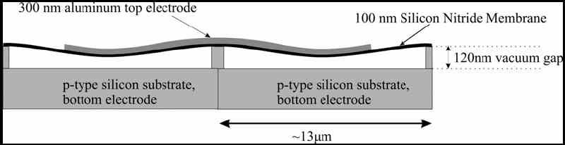
Capacitive Micromachined Ultrasonic Transducers (CMUTs) have been developed and fabricated at our department. The main goal is to use an improved version of these structures to perform medical imaging to detect unstable plaque in the coronary arteries. Unstable plaques are fatty lipid pools contained in the wall of the coronary arteries by a thin fibrous cap. A rupture of this cap can lead to an infarction. The CMUTs have a radius of 5.7?m and a center frequency of about 30MHz in air. When an RF voltage is applied in addition to a DC bias the membrane will vibrate and generate ultrasound waves. This DC bias is in the order of 30V and leads to a charge diffusion in the CMUTs. This work investigates this process in detail.
Statement of Contribution/Methods:
A heterodyne interferometer has been built in order to characterize the CMUTs. The setup can measure absolute phase and amplitudes. By using two acousto-optic modulators in the reference arm of the interferometer we can measure acoustic frequencies in the range 10kHz-1.2GHz. The results from the interferometer are supplemented with measurements from a network analyzer. The network analyzer takes the mean of all currents generated by CMUTs whereas the interferometer inspects individual CMUT elements.
Results:
The vibrating membrane in the CMUT is made of silicon nitride, which ideally is an insulator. However, we observe a charge diffusion through this membrane influencing the response of the CMUTs. There are two possible mechanisms. One is that positive charges diffuse from the bottom electrode through the silicon substrate and into the silicon nitride membrane. The other is that negative charges from the top electrode diffuses into the silicon nitride membrane. An experiment investigating the resonance frequency as a function of time indicated that the latter mechanism is dominant. Measurements from both the interferometer and the network analyzer supported this conclusion.
Discussion and Conclusions:
The measurements presented here are performed in air. Under loading conditions such as water or tissue the frequency bandwidth of the CMUT increases substantially. The charge diffusion problem may therefore not be a major problem when the CMUT is operated in water.
|
|
Finalist #5.2 (PS020-20) (3G-4):
Title: High-frequency (>100MHz) Piezoelectric PZT Film Micromachined Ultrasonic Arrays
*Dawei Wu (Presenter), *Qifa Zhou, **Changgeng Liu, **Frank Djuth, and *K Kirk Shung, *NIH Transducer Resource Center and Department of Biomedical Engineering, University of Southern California, USA,
**Geospace Research, Inc, USA. (Abstract ID: 858)
Abstract:
Background, Motivation and Objective:
High frequency (>30 MHz) ultrasonic imaging has been extensively used for imaging of the eye, blood vessel, skin and small animals. Fabrication of the transducers, which is the most critical component of the ultrsound imaging system, becomes especially challenging when very high frequency (>100 MHz) is required. Conventional lapping-and-dicing methods with bulk piezoelectric materials are no longer a viable approach. During recent years, the advance of microelectromechanical system (MEMS) methods has offered significant opportunities for miniaturized devices. This paper presents the latest development of high-frequency (>100MHz) micromachined ultrasonic linear arrays with highquality PZT thick films.
Statement of Contribution/Methods:
Both kerfless and kerfed arrays were fabricated with the PZT thick films which were prepared by spin-coating PZT composite solution. To fabricate the kerfless array, a layer of Cr/Au was patterned onto PZT film surface by using photolithographic techniques. A conductive epoxy, E-solder 3022, was used as a backing material after the silicon substrate was removed. One major problem with the kerfless arrays is their large crosstalk. To decrease the crosstalk, Inductively coupled plasma-Reactive ion etching (ICP-RIE) SF6 based dry etching was selected to etch the PZT thick films into kerfed arrays. The kerfs of the array were next filled with non conductive epoxy; the front surface of the array elements were coated with Cr/Au electrodes. E-solder was poured in as the backing material after etching away the silicon substrate.
Results:
A representative element of the kerfless array was found to have a center frequency of 120 MHz, -6 dB bandwidth of 40% and an insertion loss of around -40 dB. Its bandwidth increased to 60% after a layer of parylene was deposited as a matching layer. The etched PZT film array has a thickness of 15 ?m and etched profile angle of 75?as shown below. Charactrization of the array has been carried out. Results show great promise for this technology in fabricating linear arrays at a frequency higher than 50 MHz.
Discussion and Conclusions:
High-frequency (>100 MHz) PZT linear kerfless arrays are fabricated and tested. Preliminary results of the etched linear kerf array are promising. The results show that integrating PZT films into MEMS devices can serve as a feasible solution to high-frequency ultrasonic array fabrication.
|
|
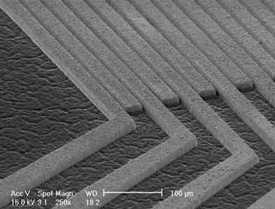
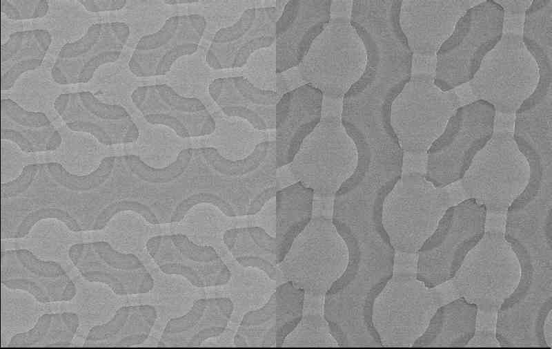
Finalist #5.3 (PS021-21):
Title: 1-D CMUT Imaging Arrays Fabricated Using a Novel Wafer Bonding Process
Andrew Logan (Presenter) and John Yeow, Systems Design Engineering, University of Waterloo, Waterloo, Ontario, Canada. (Abstract ID: 418)
Abstract:
Background, Motivation and Objective:
Capacitive micromachined ultrasonic transducers (CMUTs) are a promising alternative to conventional piezoelectric transducers for medical imaging and diagnostics.They have demonstrated image quality on par with commercial piezoelectric transducers, while the use of semiconductor fabrication technologies to manufacture ultrasonic imaging arrays has a number of advantages such as batch fabrication, reduced element lay-out constraints and the potential for onchip electronic circuit integration.
Statement of Contribution/Methods:
Here we report the fabrication and testing of 1-D CMUT arrays using a novel wafer bonding process whereby the membrane and the insulation layer are both silicon nitride. The use of a user-grown insulating membrane layer avoids the need for expensive SOI wafers, reduces parasitic capacitance, and allows more freedom in selecting the membrane thickness while also enjoying the other benefits of wafer bonding fabrication.
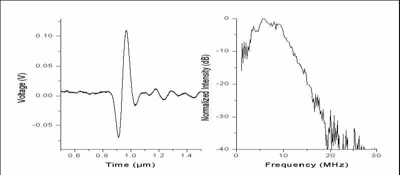
Results:
A 128x1 element array with a center frequency of 16 MHz in air and 7 MHz in immersion and a 64x1 element array with a center frequency of 40 MHz in air 25 MHz in immersion are discussed. Figure 1 is an SEM image of the 128 element device (left) and 64 element device (right). Figure 2 is the transmission pulse of the 128 element device (left) and the Fourier transform of the transmission (right). The device is biased at 100 V and a voltage spike is applied using a commercial ultrasonic pulser/receiver. Signal is detected using a commercial hydrophone. The device has a -6 dB bandwidth of 105%.
Discussion and Conclusions:
The devices discussed here are suitable for phased array imaging without grating lobes. The element dimensions are 100 ?m x 5 mm and 30 ?m x 2 mm for the 128 and 64 element devices respectively. The devices will be used for biological imaging purposes.
|
|
|
Home Contact the webmaster, Dr. Jian-yu Lu, for questions. © Copyright 2006-2008 IEEE UFFC Society