High School Teacher Outreach
Program
Monday, November 11 at Thomas Jefferson National Accelerator
Facility (JLab)
Speakers:
Abstract:
Local teachers have been invited to spend the day at Jefferson
Lab. The day will start at the Lab's auditorium (CEBAF Center)
at 8:45 with an introduction by the JLab Science Education Manager,
Jan Tyler, and by the NSS Program Chair, Nigel Lockyer. This
will be followed by the series of talks listed above. The evening
talk by Michael Levi will be open to the public.
Directions to Jefferson Lab are available at: https://www.jlab.org/user_resources/travel/maps/maps4.html.
For further information about this event, please contact Jan
Tyler via email or by phone
at 757-269-7164.
The Physics of Nuclear Medical Imaging
Bill Moses
Lawrence Berkeley National Lab
This talk describes the physics underlying Nuclear Medical
Imaging, a
group of techniques that are commonly used to diagnose and assist
in
treatment planning for cancer, heart disease, and neurological
diseases such as Alzheimer's disease and Parkinson's disease.
With
these techniques, the patient is injected with a radioactive
drug.
The drug accumulates within the body depending on the biological
nature of the drug and the disease (for example, there is a
radioactive sugar that accumulates in rapidly growing cancers).
The
drug undergoes radioactive decay and gamma rays that are emitted
by
the drug pass through the patient. These gamma rays are imaged,
and
the resulting image helps the physician diagnose and treat patients.
Two commonly types of nuclear medical imaging are known as Single
Photon Emission Computed Tomography (SPECT), where the drug that
is
used emits a single gamma ray, and Positron Emission Tomography
(PET), where the drug that is used effectively emits a pair of
back-to-back gamma rays.
The talk concentrates on the physics of nuclear medical imaging,
but
will also describe the medical motivation and the reasons for
using
nuclear medical imaging rather than other medical imaging techniques
(such as x-ray, ultrasound, or MRI). The topics discussed will
include the different radioisotopes used and the methods used
to
produce them, the physics of image formation, the underlying
principles of the PET and SPECT cameras that image the emitted
radioactivity, and the mathematics used to reconstruct images.
Solar Neutrino Astronomy: Birth After
30 Years of Labor
Josh Klein, Asst. Prof. of Physics
University of Texas, Austin, TX
Thirty years ago, Ray Davis and his colleagues made the first
attempt to study the Sun using neutrinos, particles which interact
with matter so weakly that they can travel through through the
Earth more easily than sunlight through a window pane. The Sun
produces neutrinos in the nuclear reactions which provide its
power, and the neutrinos come to us directly from the Sun's center
where the energy is produced. Davis's experiment was expected
to be a great triumph for the nuclear power theory of the Sun
and the beginning of solar neutrino astronomy, but what he found
was a surprise: the neutrinos were there, but there were not
nearly enough of them. Before being able to use neutrinos to
understand the Sun, we had to understand what was happening to
the neutrinos.
After three decades and six different experiments, we finally
believe we know the answer. The recent results from the Sudbury
Neutrino Observatory tell us that the neutrinos the Sun is producing
are changing from one type into another---all the neutrinos are
there, we just weren't looking at the right ones. And while this
solves one old problem, it helps create a new one, for it tells
us that our most fundamental theories of the behavior of the
microscopic Universe are at best incomplete. For twenty years
our model of the fundamental particles has been mapped in great
detail, but with the results from SNO and other recent neutrino
experiments, we have discovered that there is territory yet to
be explored.
With SNO's results we therefore have the creation of two new
fields, one in which we will strive to understand the newly discovered
properties of neutrinos themselves, and one in which we return
to Davis's original goal, using neutrinos to understand what
happens inside the Sun.
Nuclear and Medical
Instrumentation Development at RMD
Kanai S. Shah
Radiation Monitoring Devices, Watertown, MA 02472
Radiation Monitoring Devices (RMD), a small, high technology
company is involved in development of products based on nuclear
instrumentation for use in medical, environmental, nuclear waste
cleaning, nuclear non-proliferation and other related applications.
Activities at RMD include research and development of sensors,
instruments, and systems. Room temperature semiconductors, novel
scintillators, solid state optical detectors, new digital imaging
detectors, and multi-element electronic readout systems are all
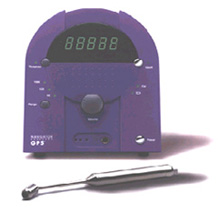
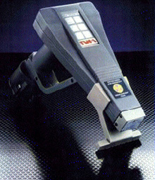
being investigated at RMD. RMD has commercialized various products
utilizing nuclear instrumentation. This includes a surgical probe
system (shown below on the left) that is used in detection of
radiolabeled tissue for detection of breast cancer. Another system
uses a room temperature semiconductor detector for detection
of lead that may be present in house-hold paint (shown below
on the right). Various other sensors and systems in research
and development stages will also be discussed. Further information
about RMD's research activities as well as its commercial products
can be obtained at RMD's website: https://www.rmdinc.com
**Special Evening Presentation - Open to the Public **
Supernova / Acceleration Probe
(SNAP): Studying the Dark Energy of the Universe
Michael Levi
Lawrence Berkeley National Laboratory
Recent measurements carried out by the Supernova Cosmology
Project (SCP) and the High-Z Supernova team have made the startling
discovery that the expansion of the universe is accelerating.
The source of this acceleration is more powerful than the gravitation
from all seen and unseen forms of matter and known energy. Michael
Levi will discuss how the Supernova / Acceleration Probe (SNAP)
Mission will provide an understanding of the mechanism driving
the acceleration of the universe.
Return to top of page
Compton scatter
imaging workshop
Thursday, November 14, 2002, 7:00 to 9:00 pm
Marriott - Norfolk Ballroom 1st Floor
Registration: none, open to all
| Chairman: |
Gary Royle, University
College London |
7:00 Compton cameras for medical imaging
Neal Clinthorne, University of Michigan
7:30 The advanced Compton telescope
Richard Kroeger, Naval Research Laboratory, Washington
8:00 Development of a high-pressure xenon electroluminescence
absorption detector with fibre readout for a Compton camera
Alexander Bolozdynya, Constellation Technology Corporation
8:15 A Novel High Resolution Compton Detector for Positron
Emission Tomography
Tumay Tumer, University of California
8:30 Compton camera question and answer session with
expert panel
Return to top of page
Workshop on the
Nuclear Radiology of Breast Cancer
Saturday and Sunday, November 16-17, 2002
Registration fee: USD $150
Registration deadline: November 1, 2002. There will be no on-site
registration.
| Organizers: |
Martin Tornai, Duke
University
Craig Levin, UCSD |
ABSTRACT
This one-and-a-half day workshop is supported in part by the
Susan G. Komen Breast Cancer Foundation and the IEEE NPSS. The
workshop will cover issues related to nuclear emission imaging
for breast cancer evaluation. Topics will include specific biological
markers, radiotracers, new instrumentation and methods designed
for breast cancer identification and localization, comparison
to conventional and other emerging breast imaging technologies,
clinical practicality issues, cost-effectiveness, industrial
perspectives, and funding opportunities
STUDENT STIPENDS
There will be five USD $500 travel grants available for graduate
students or post-doctoral fellows to attend the workshop. Interested
applicants who have made contributions to research relevant to
nuclear emission breast imaging should submit a short list of
qualifications and a one-to-two paragraph (max. one page) statement
as to why they should be awarded a grant to attend the workshop.
If applicable, the student/post-doc grant applicant may also
include the abstract and two pages of supporting data describing
their work that was submitted to the 2002 IEEE NSS/MIC meeting.
Completed travel grant applications should be submitted by e-mail
to CLEVIN@UCSD.EDU and MARTIN.TORNAI@DUKE.EDU
no later than October 18, 2002.
WORKSHOP OUTLINE
DAY ONE: Saturday, November 16, 2002, 12:30PM
- 7:30PM (including lunch and dinner):
- LUNCHEON
I. Background and Significance of Breast Cancer/Disease
- A. Biological Markers and Their Use in Breast Cancer Detection
(Margaret Huflejt, PhD)
- B. Digital Mammography (Laurie Fajardo, MD)
- II. Breast Imaging and Disease Management with Clinical Nuclear
Medicine
- A. The Role of Nuclear Medicine: Methods and Pitfalls
- (David Mankoff, MD,PhD)
- B. The Need for Nuclear Medicine Techniques for Breast Imaging
(Iraj Khalkhali, MD)
- C. Evidence-Based Breast Imaging Technology: Payer Derived
Cost-Effectiveness (Frank Papatheofanis, MD,PhD)
- III. Questions & Panel Discussion
- DINNER - Discussion
-