mic
The Medical Imaging Conference (MIC) is a unique international scientific meeting on the physics, engineering and mathematical aspects of nuclear medical imaging. The meeting will take place within the context of an increasing interest in multimodality approaches, both in terms of development of integrated multimodality imaging devices as well as in unique software developments in image reconstruction and processing, making use of synergies associated with the combination of functional and anatomical imaging modalities and beyond. In parallel, developments in radiotherapy instrumentation and associated treatment and dosimetry protocols, including the combination of imaging and radiation therapy, are continuously gaining ground. The contents of this MIC meeting will therefore reflect the expanding areas of hardware and software developments both in multimodality imaging and radiation therapy through a number of specialized topics. The MIC program format consists of oral and poster sessions. In addition, joint sessions will be held with the NSS and RTSD communities, to highlight transverse research works in specific areas. Regular sessions will be complemented by Short Courses covering all different imaging and therapy aspects.
Authors are invited to submit papers describing their original work in one of the MIC topics listed below or for the joint sessions on dosimetry, counting photon detectors / spectral CT, and hadron therapy.
The MIC program topics are listed below.
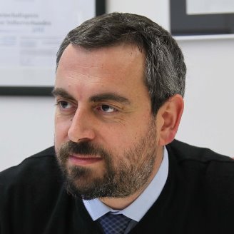
Dimitris Visvikis
MIC Program Chair
INSERM, UMR 1101, LaTIM, Brest, France
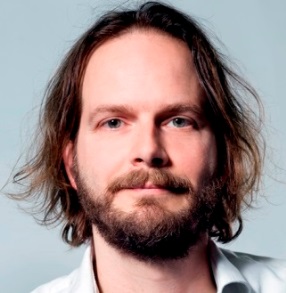
Suleman Surti
MIC Deputy Program Chair
University of Pennsylvania, Philadelphia, USA
mic topic description
Extensive research efforts are underway to better take advantage of physically available signals, either to enhance the performance of established imaging techniques (e.g., ultra-fast PET detectors), or to use new information (e.g., spectral X-ray and CT imaging). This session will target original contributions specifically addressing developments in emerging new detector materials and technologies for a broad range of applications in medical imaging, with special emphasis on recent advances in the field (e.g., ultrafast scintillators, single photon counting detectors). Moreover, presentation and evaluation of innovative detector materials/technologies that are promising but not mature enough for clinical or pre-clinical use are particularly encouraged.
Technical and performance aspects related to pre-clinical molecular imaging instrumentation, techniques and systems such as spatial resolution, sensitivity, scanning efficiency, accuracy, and reproducibility. Special emphasis is on advances in high-resolution systems that push current performance limits (e.g., sub mm micro PET, sensitivity) to explore new molecular horizons as well as multi-modality small animal (e.g., mice, rat) scanners.
The growing clinical use of nuclear medical imaging in a broad range of applications for diagnostic as well as therapy monitoring purposes is posing new demands on the imaging performance of Emission Tomography instrumentation. While basic developments in novel detector technology used in multimodal devices will be covered in New Detector Materials/Technologies for Medical Imaging / Multi-Modality Systems, the focus of this specific topic will be on complete imaging system solutions. This includes innovative developments towards significant improvements in key features (e.g., sensitivity, spatial resolution, timing resolution, …), assessment of performance and image quality of PET, SPECT instrumentation and applications of multi-modal systems specifically aiming at clinical applications. Contributions addressing advances in hardware and software solutions for seamless integration of at least two imaging modalities (X-ray, CT, optical, MRI), one of which being either PET or SPECT, are also encouraged.
This session will focus on all imaging aspects related to development of intra-operative probes (e.g., gamma probes, beta probes as well as optical and other probes using non-ionizing radiation) with special emphasis on advances for monitoring therapy, minimally invasive treatment, as well as novel portable and/or dedicated imaging systems (e.g., dedicated cardiac, brain, or breast PET or SPECT, portable CT, mobile PET-CT), that open new opportunities in bedside imaging (e.g., intensive care units, in operating rooms, in-room imaging during treatment).
Original work pertaining to ionizing medical imaging modalities that do not rely on the visualization of single photon or positron emitting tracers (e.g., X-ray, computed tomography) will be the focus of this session. New hardware and software addressing current issues of transmission imaging, including improved image reconstruction techniques, 4D imaging, multi-spectral CT developments for both clinical and preclinical applications are particularly welcomed.
Original work pertaining to non-ionizing medical imaging modalities (e.g., magnetic resonance, optical, ultrasound imaging) should be submitted to this topic. Novel work using advanced imaging techniques that can be applied across modalities (e.g., sparse sampling, acceleration techniques) is particularly encouraged.
Quantitative medical imaging is affected by physical factors (e.g., scatter in ionizing imaging, noise, limited spatial resolution) as well as factors associated with patient physiology (e.g., respiratory and cardiac motion) that limit the accuracy that can be achieved in-vivo. This track pertains to novel techniques for compensating for the physical and other technical factors as well as the physiological factors affecting quantitation with special emphasis on recent advances in quantitative imaging.
This session will emphasize the development of novel approaches for the assessment of image quality as well as studies assessing existing algorithms for improved image qualitative and quantitative accuracy (including image reconstruction and post-reconstruction processing methodologies).
Novel work in the area of image reconstruction for any/all medical imaging modalities (e.g., SPECT, PET, CT, MRI, OCT) is encouraged for submission. Both analytical and model based iterative approaches are welcomed. Novel approaches for accelerated reconstruction, sparse sampling, model based reconstruction, etc. as well as the evaluation of the performance of these approaches are also welcomed.
The overwhelming increase of multi-modal information at the detector and image level makes it crucial to advance research for the development of algorithmic and automated workflows of signal and image processing that can reduce human intervention while preserving its experienced judgment. This track is devoted to original work in signal and image processing, as well as novel evaluation metrics. In particular, innovative contributions in evaluation methodology paving the way for reliable and cost-effective assessment of medical image quality is welcomed.
Kinetic modeling of physiological processes (e.g., radiotracers kinetics) is essential to understanding the pharmacokinetics of novel imaging and therapeutic agents which can be captured in parametric imaging. This track focuses on all aspects related to parametric imaging and tracer kinetic modeling, with special emphasis on new tracers and modeling techniques, as they pertain to pre-clinical and clinical imaging.
Ongoing research efforts aiming at developing increasingly complex instrumentation for medical imaging rely on the use of sophisticated simulation tools to reproduce with high fidelity the main aspects of the image formation chain, including interactions in the patient and detector and system assembly. This topic focuses on original validation and development of simulation tools aiming to support hardware and software developments in single or multi-modalilty imaging systems, as well as their application to medical imaging in diagnostics and therapy, with special emphasis on nuclear medicine. In particular, we welcome innovative contributions addressing crucial modeling aspects in terms of computational efficiency and accuracy in reproducing the physical interactions which affect the performances of the medical imaging systems.
Recent advances in the precision of the delivery of photon and hadron beams has yielded increasing developments in the field of image guided radiotherapy. This topic will include innovative research for the integration of imaging at the treatment site, as well as emerging techniques of in-vivo range verification in hadron therapy. Software solutions allowing an improved dosimetry for external beam and intra-operative radiotherapy applications are included in this topic. Novel applications of existing technologies or innovative solutions offering new image guidance possibilities in photon and ion beam therapy are welcomed.
mic plenary speakers
Redefining optical imaging with Multi-Spectral Optoacoustic Tomography
Prof. Vasilis Ntziachristos, Technische Universität München, Germany
biography

Prof. Ntziachristos´s area of research encompasses the development of new methods and devices for biological and medical imaging focusing on innovative non-invasive approaches that visualize previously unseen physiological and molecular processes in tissues. His research also aims to translate these methods to advance biological dis-covery, accelerate drug development and offer new efficient methods for diagnostics and theranostics. Prof. Ntziachristos studied electrical engineering at Aristotle University in Thessaloniki, Greece and received his Master’s and Doctorate degrees from the Bioengineering Department of the University of Pennsylvania. He served as assistant professor and director of the Laboratory for Bio-Optics and Molecular Imaging at Harvard University and Massachusetts General Hospital. His current professorial chair is closely linked with the Institute for Biological and Medical Imaging at the Helmholtz Zentrum München, of which Prof. Ntziachristos is director.
abstract
Optical imaging is unequivocally the most versatile and widely used visualization modality in the life sciences. Yet it has been significantly limited by photon scattering, which complicates imaging beyond a few hundred microns. For the past few years, there has been an emergence of powerful new optical and optoacoustic imaging methods that can offer high resolution imaging beyond the penetration limits of microscopic methods. The talk discusses progress in multi-spectral opto-acoustic tomography (MSOT) that brings unprecedented optical imaging performance in visualizing anatomical, physiological and molecular imaging biomarkers. Some of the attractive features of the method is the ability to offer 10-100 microns resolution through several millimetres to centimetres of tissue and real-time imaging. We demonstrate implementations in the time and frequency domain, showcase how it is possible to accurately solve fluence and spectral coloring issues for yielding quantitative measurements of tissue oxygenation and hypoxia and demonstrate applications in resolving inflammation, metabolism, angiogenesis in label free mode. In parallel we summarize progress with clinical systems that offer to change readings of disease in-vivo and discuss complementarity with ultrasound imaging, fluorescence imaging and other modalities. Finally the talk offers insights into new miniaturized detection methods based on ultrasound detection using optical fibers, which could be used for minimally invasive applications.
Radiomics – Mining images to make better decisions in cancer therapy
Prof. Andre Dekker, MAASTRO Clinic, The Netherlands
biography

Prof. Andre Dekker is a board-certified medical physicist at MAASTRO Clinic, Maastricht, The Netherlands since 2005. He has been the head of Medical Physics until 2009 and then led for numerous years the department of Information and Services that manages medical informatics and ICT. He is now responsible for all Research and Education activities at the hospital. He was appointed as a full professor at Maastricht University in 2015 where he holds the chair “Clinical Data Science”. From 2008, he started and is the principal investigator of the GROW-Maastricht University research division of MAASTRO Knowledge Engineering. His research focuses on two main themes: 1) global data sharing infrastructures; 2) machine learning on this data for decision support systems. The main scientific breakthrough has been the development of a Semantic Web and ontology based data sharing and distributed learning infrastructure that does not require data to leave the hospital. This has reduced many of the ethical and other barriers to share data. Prof. Dekker has authored over 90 publications (h-index 37) in peer reviewed journals covering informatics, imaging, radiotherapy, tissue optics and heart disease and holds multiple awarded patents. He has held visiting scientist appointments at the Christie Hospital NHS trust; University of Sydney Australia; Liverpool and Macarthur Cancer therapy centres Australia; Illawarra Shoalhaven Local Health District Australia; Universita Cattolica Del Sacro Cuore, Italy; Radiation Therapy Oncology Group, USA, Varian Medical Systems, USA and the Princess Margaret Hospital in Canada.
abstract
The rise of radiomics, the high-throughput mining of quantitative image features from (standard-of-care) medical imaging for knowledge extraction and application within clinical decision support systems (please watch the animation: https://youtu.be/Tq980GEVP0Y) to improve diagnostic, prognostic, and predictive accuracy, has significant and substantial implications for the medical community. Radiomic analysis exploits sophisticated image analysis tools and the exponential growth of medical imaging data to develop and validate powerful image-based signatures/models. This lecture describes the process of radiomics, its pitfalls, challenges, opportunities, and its capacity to improve clinical decision making (presently primarily in the care of patients with cancer, however, all imaged patients may benefit from quantitative radiology). Finally, the field of radiomics is emerging rapidly, however, the field lacks standardized evaluation of both the scientific integrity and the clinical significance of the numerous published radiomics investigations resulting from this growth. The lecture will discuss the need for rigorous evaluation criteria and reporting guidelines in order for radiomics to mature as a discipline.
mic student awards
first award (oral presentation)
Chen-Ming Chang, Stanford University, USA
M08-7: Time-over-Threshold for Pulse Shape Discrimination in a Time-of-flight/Depth-of-Interaction Phoswich PET Detector
second award (poster presentation)
Audrey Corbeil Therrien, Université de Sherbrooke, Sherbrooke and Physics Department, CERN
M13B-4: Energy Discrimination Using First Emitted Photon Timestamps: an Exploratory Study
third award (oral presentation)
Anna Turco, KU Leuven, Leven, Belgium
M19-2: Ex Vivo and in Vivo Study Evaluating Edge-Preserving and Anatomical Priors for Partial Volume Correction in Cardiac PET
fourth award (poster presentation)
Cameron Miller, University of Michigan, Ann Arbor and Loma Linda University, Loma Linda
M10C-10: Scintillator-Based Measurement of off-Axis Neutron and Photon Dose Rates During Proton Therapy

