

MIC
The IEEE Medical Imaging Conference (MIC) is the leading international scientific medical imaging meeting bringing together a broad community interested in the physics, engineering, and mathematical aspects of medical imaging. As the field rapidly evolves towards interdisciplinary, multi-modality approaches, the topics covered in the conference range from nuclear medicine (SPECT and PET) to X-ray, CT, optical, MR imaging, and their combination. In addition to this broad portfolio, Joint NSS-MIC sessions will cover hardware topics of interest to both communities and Short Courses will cover specialized timely topics. Authors are invited to submit papers describing original, previously unpublished work pertaining to the topics listed here.
Georges El-Fakhri
MIC Program Chair
Harvard Medical School, USA
Katia Parodi
MIC Deputy Program Chair
Ludwig Maximilians University, Germany
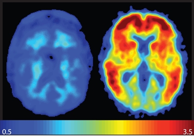
 Quantitative medical imaging is affected by physical factors (e.g., scatter in ionizing imaging, noise, limited spatial resolution) as well as factors
associated with patient physiology (e.g., respiratory and cardiac motion) that limit the accuracy that can be achieved in-vivo. This track pertains to
novel techniques for compensating for the physical
and other technical factors as well as the physiological factors affecting quantitation with special emphasis on recent advances in quantitative imaging.
Quantitative medical imaging is affected by physical factors (e.g., scatter in ionizing imaging, noise, limited spatial resolution) as well as factors
associated with patient physiology (e.g., respiratory and cardiac motion) that limit the accuracy that can be achieved in-vivo. This track pertains to
novel techniques for compensating for the physical
and other technical factors as well as the physiological factors affecting quantitation with special emphasis on recent advances in quantitative imaging.
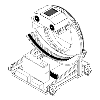
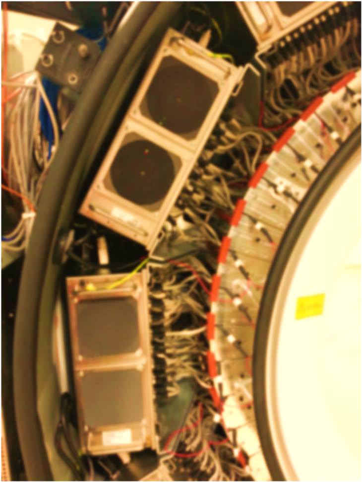 The growing clinical use of nuclear medicine imaging in a broad range of applications in diagnostic imaging as well as therapy and
theranostics is posing new demands on the imaging performances of Emission Tomography instrumentation. While basic developments in novel detector
technology and multimodal systems will be covered in
The growing clinical use of nuclear medicine imaging in a broad range of applications in diagnostic imaging as well as therapy and
theranostics is posing new demands on the imaging performances of Emission Tomography instrumentation. While basic developments in novel detector
technology and multimodal systems will be covered in
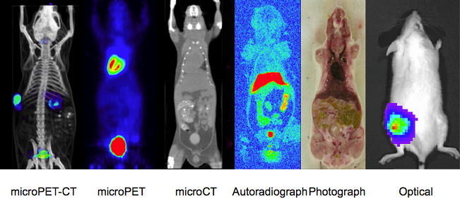 Technical and performance aspects related to pre-clinical molecular imaging instrumentation, techniques and systems such as spatial resolution,
sensitivity, scanning efficiency, accuracy, and reproducibility. Special emphasis is on advances in high-resolution systems that push current
performance limits (e.g., sub mm micro PET, sensitivity)
to explore new molecular horizons as well as multi-modality small animal (e.g., mice, rat) scanners.
Technical and performance aspects related to pre-clinical molecular imaging instrumentation, techniques and systems such as spatial resolution,
sensitivity, scanning efficiency, accuracy, and reproducibility. Special emphasis is on advances in high-resolution systems that push current
performance limits (e.g., sub mm micro PET, sensitivity)
to explore new molecular horizons as well as multi-modality small animal (e.g., mice, rat) scanners.
 Novel work in the area of image reconstruction for any/all medical imaging modalities (e.g., SPECT, PET, CT, MRI, OCT)
is encouraged for submission. Both analytical and model based iterative approaches are welcomed. Novel approaches for
accelerated reconstruction, sparse sampling, model based reconstruction,
etc. as well as the evaluation of the performance of these approaches are also welcomed.
Novel work in the area of image reconstruction for any/all medical imaging modalities (e.g., SPECT, PET, CT, MRI, OCT)
is encouraged for submission. Both analytical and model based iterative approaches are welcomed. Novel approaches for
accelerated reconstruction, sparse sampling, model based reconstruction,
etc. as well as the evaluation of the performance of these approaches are also welcomed.
 Recent advances in the precision of the delivery of photon and hadron beams has yielded increasing developments in the development of
unconventional approaches to image guided radiotherapy. This track focuses on innovative research for the integration of imaging at the
treatment site, as well as emerging techniques of in-vivo range verification in hadron therapy. Novel applications of existing technologies
or innovative solutions offering new image guidance possibilities in photon and ion beam therapy are welcomed.
Recent advances in the precision of the delivery of photon and hadron beams has yielded increasing developments in the development of
unconventional approaches to image guided radiotherapy. This track focuses on innovative research for the integration of imaging at the
treatment site, as well as emerging techniques of in-vivo range verification in hadron therapy. Novel applications of existing technologies
or innovative solutions offering new image guidance possibilities in photon and ion beam therapy are welcomed.
 All aspects related to intra-operative probes (e.g., gamma probes, beta probes as well as optical and other probes using
non-ionizing radiation) with special emphasis on advances for monitoring therapy, minimally invasive treatment, as well
as novel portable and/or dedicated imaging systems (e.g., portable CT, mobile PET-CT) that open new opportunities in bedside imaging
(e.g., intensive care units, in the surgery theater, in room imaging during treatment).
All aspects related to intra-operative probes (e.g., gamma probes, beta probes as well as optical and other probes using
non-ionizing radiation) with special emphasis on advances for monitoring therapy, minimally invasive treatment, as well
as novel portable and/or dedicated imaging systems (e.g., portable CT, mobile PET-CT) that open new opportunities in bedside imaging
(e.g., intensive care units, in the surgery theater, in room imaging during treatment).
 The growing inter-disciplinarity of biomedical engineering has been accompanied by an increasing effort to combine different
imaging information not only virtually on a computer, but also via hardware integration of different imaging technologies in
multi-modal systems. While efforts related to pre-clinical small animal imaging will be covered in
The growing inter-disciplinarity of biomedical engineering has been accompanied by an increasing effort to combine different
imaging information not only virtually on a computer, but also via hardware integration of different imaging technologies in
multi-modal systems. While efforts related to pre-clinical small animal imaging will be covered in
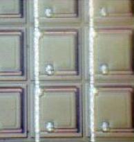 Extensive research efforts are underway to better take advantage of physically available signals, either to enhance
the performances of established imaging techniques (e.g., ultra-fast PET detectors), or to use new information
(e.g., spectral X-ray and CT imaging). This session will target original contributions specifically addressing developments
in emerging new detector materials and technologies for a broad range of applications in medical imaging, with special emphasis
on recent advances in the field (e.g., ultrafast scintillators, single photon counting detectors). Moreover, presentation and
evaluation of innovative detector
materials/technologies that are promising but not mature enough for clinical are particularly encouraged.
Extensive research efforts are underway to better take advantage of physically available signals, either to enhance
the performances of established imaging techniques (e.g., ultra-fast PET detectors), or to use new information
(e.g., spectral X-ray and CT imaging). This session will target original contributions specifically addressing developments
in emerging new detector materials and technologies for a broad range of applications in medical imaging, with special emphasis
on recent advances in the field (e.g., ultrafast scintillators, single photon counting detectors). Moreover, presentation and
evaluation of innovative detector
materials/technologies that are promising but not mature enough for clinical are particularly encouraged.
 Original work pertaining to ionizing medical imaging modalities that do not rely on the visualization of single photon or positron
emitting tracers both ionizing (e.g., X-ray, computed tomography) as well as non ionizing modalities (e.g., magnetic resonance,
optical, ultrasound imaging) is encouraged to be submitted to the conference. Novel work using advanced imaging techniques
that can be applied across modalities
(e.g., sparse sampling, acceleration techniques) is particularly encouraged.
Original work pertaining to ionizing medical imaging modalities that do not rely on the visualization of single photon or positron
emitting tracers both ionizing (e.g., X-ray, computed tomography) as well as non ionizing modalities (e.g., magnetic resonance,
optical, ultrasound imaging) is encouraged to be submitted to the conference. Novel work using advanced imaging techniques
that can be applied across modalities
(e.g., sparse sampling, acceleration techniques) is particularly encouraged.
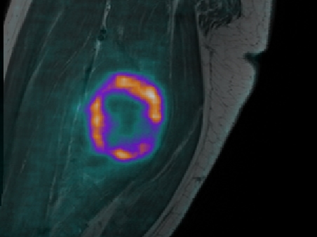
 Kinetic modeling of physiological processes (e.g., radiotracers kinetics) is essential to understanding the pharmacokinetics
of novel imaging and therapeutic agents which can be captured in parametric imaging. This track focuses on all aspects related
to parametric imaging and tracer kinetic modeling, with special
emphasis on new tracers and modeling techniques, as they pertain to pre-clinical and clinical imaging.
Kinetic modeling of physiological processes (e.g., radiotracers kinetics) is essential to understanding the pharmacokinetics
of novel imaging and therapeutic agents which can be captured in parametric imaging. This track focuses on all aspects related
to parametric imaging and tracer kinetic modeling, with special
emphasis on new tracers and modeling techniques, as they pertain to pre-clinical and clinical imaging.
 The overwhelming increase of multi-modal information at the detector and image level makes it crucial to advance research
for the development of algorithmic and automated workflows of signal and image processing that can reduce human intervention
while preserving its experienced judgment. This track is devoted to original work in signal and image processing, as well as
novel evaluation metrics. In particular, innovative contributions in evaluation methodology paving
the way for reliable and cost-effective assessment of medical image quality is welcomed.
The overwhelming increase of multi-modal information at the detector and image level makes it crucial to advance research
for the development of algorithmic and automated workflows of signal and image processing that can reduce human intervention
while preserving its experienced judgment. This track is devoted to original work in signal and image processing, as well as
novel evaluation metrics. In particular, innovative contributions in evaluation methodology paving
the way for reliable and cost-effective assessment of medical image quality is welcomed.
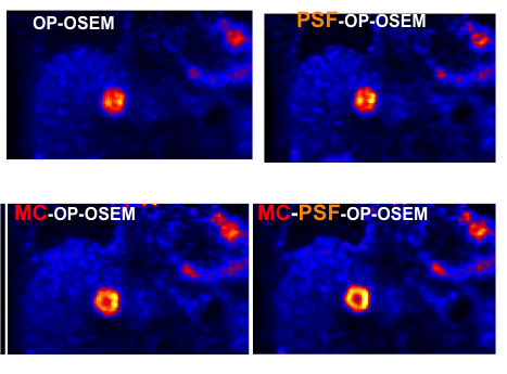
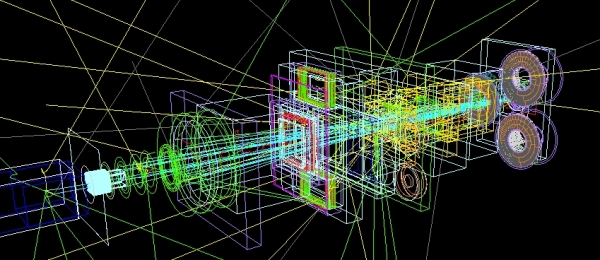
 Ongoing research efforts aiming at developing increasingly complex instrumentation for medical imaging rely on the use of
sophisticated simulation tools to reproduce with high fidelity the main aspects of the image formation chain, including interactions
in the patient and detector and system assembly. This track focuses on original validation and development of simulation tools
aiming to support hardware and software developments in single or multi-modalilty imaging systems, as well as their application
to medical imaging in diagnostics and therapy, with special emphasis on nuclear medicine. In particular, we welcome innovative
contributions addressing crucial modeling aspects in terms of computational efficiency and accuracy in reproducing
the physical interactions which affect the performances of the medical imaging systems.
Ongoing research efforts aiming at developing increasingly complex instrumentation for medical imaging rely on the use of
sophisticated simulation tools to reproduce with high fidelity the main aspects of the image formation chain, including interactions
in the patient and detector and system assembly. This track focuses on original validation and development of simulation tools
aiming to support hardware and software developments in single or multi-modalilty imaging systems, as well as their application
to medical imaging in diagnostics and therapy, with special emphasis on nuclear medicine. In particular, we welcome innovative
contributions addressing crucial modeling aspects in terms of computational efficiency and accuracy in reproducing
the physical interactions which affect the performances of the medical imaging systems.