MIC Plenary Speakers
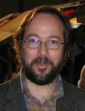
M01-1: Exploring Human Brain Architecture, Van Wedeen
Wednesday, 12 November, 8:00-9:00

Abstract
Neuroscientists have been trying to unlock the structure of the brain to unravel mysteries of brain development and evolution, and help link neurological and psychiatric disorders to abnormalities in brain structure that occur in diseases like Alzheimer’s and Parkinson’s. But the long-held idea that the brain is a set of wires criss-crossing more or less at random has never quite fit with the simplicity and efficiency of natural selection.
In this lecture, we will explore brain structure using a mapping technique called diffusion spectrum MRI. Our results reveal a fiber architecture of the brain where each pathway is one point in a three-dimensional grid not an isolated network that connects and disconnects at will with distinct left-right, up-down and front-back patterning shown in three axes, much like a three-dimensional street map. Astonishing complexity can arise from a seductively simple underlying structure that follows a few simple rules and repeats them over and over. In fact, if you straighten out the folds in the brain it turns into a three-dimensional tapestry of interwoven fibers running at 90 degrees to each another. These sheets are also arranged at right angles to one another, completing a three-dimensional grid.
We hypothesize that brain fibers grow during embryonic development following a simple set of rules. Biochemical signals determine which direction the fibers grow, much like how a city grid layout makes it easy to give directions to a destination. This simple structure might explain how complex brains evolved. If the brain were organized like a tangle of spaghetti, it would be difficult to see how mutations could lead to incremental changes in connectivity on which natural selection could act. The very simplicity of this grid structure is the reason why it can accommodate the random, gradual changes of evolution. It's easier for a simple structure to change and adapt, whether we're talking about the big changes that occur across evolution or the changes that can occur during an individual's lifetime – both the normal neuroplasticity associated with development and learning or the damage that results from injury or disease. A simple grid structure makes both evolutionary and developmental sense.
Biography
Van Wedeen MD is an Associate Professor of Radiology and Assistant in Neuroscience at MGH. A Westinghouse Scholar, he earned his BA in mathematics at Harvard College in 1977 and MD at Albert Einstein. After residency in medicine at Columbia University Roosevelt Hosp, he came to MGH in 1983 as a Fellow in MRI. His research expands the interface between biomedical imaging and mathematics. Dr Wedeen and colleagues are credited with creating the first MR angiography, published in Science in 1985. After a fellowship in Nuclear Medicine at MGH in 1990, he and colleagues developed MRI of myocardial fiber architecture and function. Turning to brain imaging, Dr Wedeen proposed the MRI of brain pathways - tractography - in 1995 and then "high angular resolution diffusion MRI" in 2000. These methods became the basis for the NIH Blueprint Human Connectome Project (HCP), of which he is now a PI. As Director of Connectomics at the Martinos Center in 2009, he leads a team of researchers to create a next-generation MRI scanner purpose-built for brain connectivity.
Recently, Dr. Wedeen's team have discovered a geometric organization of brain pathways - a 3D grid - reported in Science in 2012. His work made the cover of the National Geographic in 2013. Its implications for science and imaging, particularly for mental health, are now a focus of active research. Dr. Wedeen was elected a Fellow of the ISMRM in 2007.
M01-2: Sub mm PET would change our understanding of life and disease
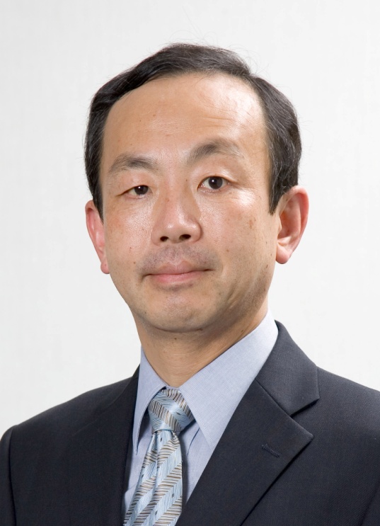
Yasuhisa Fujibayashi
Wednesday, 12 November, 9:00-10:00

Abstract
Positron emission tomography (PET) is a powerful tool in molecular imaging research, and only PET probes have allowed us to visualize molecular function of endogenous proteins such as receptors, enzymes, transporters, etc. in human. Recent progress in optical imaging probes and autoradiography probes make tissue/cellular level protein expression imaging possible. In these high resolution images (<1 mm), “heterogenous” distribution of probes could be visualized in “homogenous” PET images.
Hypoxic tumor is resistant to radiation therapy and detection of hypoxic region is a key for the prognosis of therapy. Microscopic observation revealed that hypoxic region is a patchy mixture of high vasculature surrounded by proliferating cell layer, hypoxic cell layer then necrosis, and distance between blood vessel and necrosis is c.a. 0.3 – 0.5 mm. However, in PET study, we try to find a “hypoxic region” next to “necrosis region”, using 5 – 8 mm resolution images.
Recently, a new Tau imaging agent, PBB3, has been developed. PBB3 is a phosphor and also can be labeled with C-11, so that PET, autoradiography as well as fluorescence images of PBB can be obtained. In PET, Tau accumulation was found in hippocampus region, but autoradiography and fluorescence images indicated sub-regional difference of PBB accumulation within hippocampus. To clarify the cause of disease and precise diagnosis of Tau-related diseases, sub-regional data is considered to be essential.
Inconsistency in the interpretation of PET images brings a limitation in our understanding of life and disease, even though PET is the only modality which can visualize molecular function of endogenous proteins in living human. Progress in PET technology has improved the resolution of PET for many years. Supra-high resolution PET (<1 mm) would be able to visualize such sub-regional differences in living human, and it would bring more precise understanding of life and disease.
Biography
Dr.Fujibayashi received the B.S., M.S. and Ph.D. degrees in Pharmaceutical Sciences from Kyoto University, Japan,in 1978, 1980 and 1986, respectively. He also received D.Med.Sci. degree in Medical Sciences from Kyoto University in 1995. He was appointed as Assistant Professor at Kyoto University in 1983, then Associate Professor in 1993. He moved to University of Fukui as Professor of Molecular Imaging in 1999, then co-appointed as Director of Biomedical Imaging Research Center. In 2010, he moved to National Institute of Radiological Sciences, Japan, as Director of Molecular Imaging Center. He published more than 200 peer-reviewed papers related to nuclear medicine imaging, and chaired Society for Molecular Imaging (WMIS, former SMI), Society of Radiopharmaceutical Sciences (SRS), and Japanese Society for Molecular Imaging, as President.


