Refresher Course Program

In many fields of interest, there are numerous possibilities for enhanced creativity and conceptual idea development, as well as advancement through cross-fertilization of skills and knowledge. Furthermore, broadened knowledge of related technologies helps to open new avenues of consideration in the search for novel and unique solutions to difficult problems.

All of this idealistic reasoning and rationalization aside, curiosity compels most of us to learn new things from an almost an endless list of motivations. Being interested in satisfying this thirst for knowledge, we plan to hold a series of Refresher Courses to provide the fundamental information and background in fields and technologies of particular (personal or professional) interest. We anticipate that these courses will stimulate the curiosity and interest of attendees from all three programs.


The Refresher Courses will be free for all attendees, will be about one hour in length (topic-dependent), and will take place during the two-hour lunch break (12:00 – 14:00) such that you can attend these courses and also be able to get lunch.


Please check back often to see what offerings we have for you to experience.
Course Description

Fast waveform digitizing in nuclear and particle physics is traditionally done with flash ADCs. These devices however hit their limits in resolution and power consumption when it comes to sampling rates far beyond the Giga-sample per second range (GSPS). An alternative for non-periodic signals are Switched Capacitor Arrays (SCA) that store an analog waveform in a series of capacitors, which are then digitized after a trigger at much lower speed. While these chips have been used for two decades in particle physics, the recent improvements in CMOS technology allows for designs with resolutions of 12 bits, sampling speeds beyond 10 GSPS and power consumptions of a few tens of mW per channel. Putting many channels on a single chip makes it possible to build data acquisition systems with several thousand channels at reasonable costs, space and power requirements. Obtaining the waveforms of particle detectors at high resolution allows excellent timing measurements down below one pico-seconds, doing particle discrimination and efficient pile-up rejection.

This refresher course covers the basic principles of SCAs, gives an overview of currently available chips and introduces advanced waveform processing techniques used in particle physics and gamma-ray astronomy. Experiences from the MEG experiment with 3000 SCA channels are reported. It finishes with an outlook for new chips currently under design and how they can be used in future experiments.


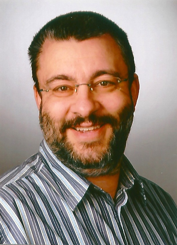
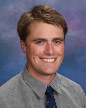
Instructor: Stefan Ritt, Paul Scherrer Institute, Switzerland

Stefan Ritt is staff member at the Paul Scherrer Institute, Switzerland, and head of the muon physics group. He received his Ph.D. in physics in 1993 from the University of Karlsruhe in Germany, after which he joined the University of Virginia, Charlottesville as a Research Scientist. His research interests are in the field of fundamental particle physics experiments and their instrumentation. He is one of the main authors of the MIDAS DAQ system, wrote the ELOG electronic logbook and co-developed the DRS switched capacitor array chip. He is author and co-author of about 50 publications, holds two patents and has been an IEEE member since 2007 and senior member since 2011.

Course Description

In recent years, a new trend in high-performance computing has evolved, which makes use of commodity graphics processors (GPUs) for massively parallel computing tasks. The increase of the processing power of GPUs, driven by high-end computer gaming, can easily be put to use for other CPU-intensive tasks. Off-the-shelf systems providing multiple TeraFlops of processing power are available at commodity price levels today.

This refresher course aims to provide a (re-) introduction to the CUDA technology, which is NVIDIA's framework for GPU programming. We will start with a few examples, which show how easy it is to go from C or C++ on the CPU to a CUDA program on GPUs. I will demonstrate some common pitfalls and show how to avoid them, and introduce some common programming techniques to produce efficient GPU code.


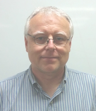
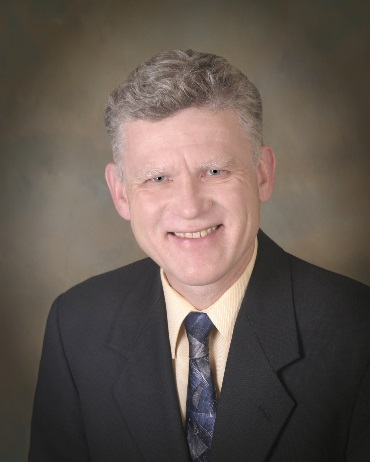
Instructor: Martin Purschke, Brookhaven National Laboratory, USA

Martin Purschke is a staff physicist at the Brookhaven National Laboratory located on Long Island, NY. He received his Ph.D. in Physics from the University of Muenster in Germany in 1990, and spent a number of years with the Heavy-Ion program at the CERN-SPS before taking a position at BNL. He is the Data Acquisition Coordinator of the PHENIX Experiment at the Relativistic Heavy Ion Collider, and has written substantial portions of the online and offline software in use in the PHENIX experiment. He is also a member in the RatCAP Pet-Imaging project at BNL. Most of his CUDA programming takes place in the framework of the medical image reconstruction for the PET detectors.

Course Description

Python is an increasingly popular scientific computing programming language, offering an easy-to-learn, versatile interface that glues together work from many other languages well. Furthermore, it is supported by a vibrant open source community. While the Python standard library is often touted for being “batteries-included”, the scientific Python environment is even richer, with many powerful tools and packages to enhance the scientific computing workflow.

In this one-hour course, we will introduce and refresh participants to Pythonic practices from the perspective of a researcher with a C or C++ background. We will cover:

  1. creating a reproducible computational environment with Docker,
  2. interactive analysis and literate programming with the IPython shell and the IPython Notebook,
  3. a brief survey of the fundamental scientific Python packages numpy, matplotlib, scipy, sympy, and pandas,
  4. writing efficient, compiled C/Python hybrid code with Cython, and
  5. wrapping C and C++ libraries in Python with XDress.

By the end of the course, participants should be more effective computational researchers through an interactive introduction into modern scientific Python practices. This is a hands-on course that requires a laptop and active participation!


Instructor: Matthew McCormick, Kitware, Inc., USA

Matthew McCormick is a medical imaging researcher working at Kitware, Inc. His research interests include medical image registration and ultrasound imaging. Matt is an active member of scientific open source software efforts such as the InsightToolkit and scientific Python communities.

Course Description

In the last 15 years, significant advances in algorithm research has been made, resulting in a rapidly growing inventory of analytic and optimization-based algorithms based upon respective data models and reconstruction programs. The presentation will provide a synopsis of some of the recent advances and issues in algorithm research for CT imaging. I will first contrast the development chains of analytic and optimization-based algorithms, their conceptual analogy/difference, and their distinctive implications for practical applications. With such a contrast analysis in place, I will then focus the discussion specifically on the development of optimization-based reconstruction because it has recently attracted a significantly increased level of research effort, (which is likely to remain, or be intensified even further, in the foreseeable future.) As a result of the effort, a large number of optimization-based algorithms have been proposed. However, realization and evaluation of the potential of the algorithm advances in real-data studies remain a highly challenging problem. Following an illustration of the design chain of optimization-based reconstruction, including reconstruction program and algorithm, I will discuss the application of an optimization-based reconstruction approach to reconstructing images in various CT imaging problems of practical implications. In particular, using a series of real CT-data examples, I will elucidate insights into, and guidance for, tailoring optimization-based reconstructions to possibly improving current applications, and to enabling the development of new imaging systems and/or applications, in medicine, security imaging, and other areas. I will also touch upon seemingly confusing issues concerning, e.g., Nyquist sampling theorem and compressive sensing (CS), CS and CT imaging, utility-based design of system/reconstruction, and evaluation paradigm for system/reconstruction development. If time allows, I will discuss potential implications of optimization-based reconstruction for, e.g., PET and MRI.


Instructor: X. Pan

Course Description

The accuracy of radiation therapy heavily relies on accurate knowledge of the distribution of heterogeneous tissues at the time of beam delivery. Imaging for treatment planning is used to determine the distribution of electron density, which is needed for photon and electron therapy, or stopping power, in the case of proton and ion therapy in 3D or, for moving targets such as lung tumors, in 4D. For pretreatment, intra-treatment, and post-treatment verification, additional imaging can be applied in the treatment room. This course will give an overview or refresh the knowledge on the technology and reconstruction techniques associated with common and evolving imaging modalities used for treatment planning and verification in radiotherapy. We will cover the following topics:

  1. imaging for treatment planning of photon and charged particle therapy,
  2. imaging and image registration for tumor and normal tissue segmentation,
  3. imaging modalities for pre-, intra- and post-treatment verification of patient position and dose delivery,
  4. challenges of charged particle beam therapy in predicting range and scattering, and
  5. the future of image-guided, adaptive radiotherapy.

After this course, participants should be familiar with all aspects of imaging for radiotherapy. This course is of interest for detector scientists as well as for medical physicists and engineers that want to refresh their knowledge of this topic or explore new areas of applying there knowledge in imaging detectors, image reconstruction and registration, or computer science and engineering and fast data processing.


Instructor: Reinhard Schulte, Loma Linda University Medical Center, USA

Reinhard Schulte is Professor in the School of Medicine at Loma Linda University, and works as Translational Researcher on proton- and ion-therapy-related technology and clinical protocols. He holds a graduate degree in Physics (Diploma) and a doctoral degree in Medicine (Dr. med., summa cum laude). He is currently Principal Investigator on an NIH-funded project to develop proton CT and participates in two large European Research Consortia related to proton therapy research. Dr. Schulte also has 25 years of experience in clinical proton therapy and is a licensed physician and board-certified radiation oncologist.

Course Description

This refresher course will provide a high-level overview of kinetic analysis in dynamic medical imaging with emphasis on the use of radiolabeled probes for PET and SPECT. We will cover basic concepts in pharmacokinetic modeling including the tracer principle, types of molecular interactions experienced by the probe, and mathematical representation of those interactions to construct a compartmental model. Specific applications will highlight studies of blood flow, metabolism, and receptor-ligand binding with discussion of the outcome parameters that can be identified from each approach. The use of non-invasive input functions and simplified modeling techniques will be presented. We will conclude by discussing current applications and areas of investigation in the field, including the use of kinetic modeling to study physiological interventions and approaches to facilitate basic research and improve clinical procedures.


Instructor: Marc Normandin, Massachusetts General Hospital, Harvard Medical School, USA

Course Description

This refresher course will be focused on one of the central components of modern emission tomography (ET) instrumentations (PET and SPECT) – gamma ray imaging detectors. It will provide an overview of the basic principle, current status and future trends for sensor development for ET applications, especially for combining PET and SPECT with other imaging modalities, such as X-ray CT and MRI.

We will start with a brief introduction to PET and SPECT instrumentations and their applications, and the need for gamma ray detectors that could precisely determine the location, energy, timing and (ideally) the type of gamma ray interactions underlying each individual event. These will be followed by discussions on basic principles and readout electronics behind different types of gamma ray detectors, such as scintillation detectors and semiconductor detectors. We will also compare several types of gamma ray detectors for PET and/or SPECT imaging, and discuss the physical factors that limit their intrinsic performances. This short course will be concluded by discussing some of the current trends in developing advanced gamma ray detectors for future emission tomography systems, and the major technical challenges involved in these efforts.


Instructor: Ling-Jian Meng, University of Illinois at Urbana-Champaign, USA

Course Description

Just 30 years ago Magnetic Resonance Imaging (MRI) was only available at prominent research institutions, while now it has become an indispensable clinical tool for medical practitioners, scientists, and engineers in thousands of institutions around the world. It is well known for its innocuousness relative to other imaging modalities, but lesser known is its wide breadth of imaging contrast. Fervent development over the years have led to the expansion of MRI into dozens of sub-modalities: it can now be used to discriminate anatomy, morphology, physiology, vascularity, cellularity, metabolism, functional connectivity/activity, perfusion, diffusion, susceptibility, elasticity, relaxivity, temperature, flow, tissue/current density, and corticality. The first half of this lecture will be dedicated to outlining the fundamentals of MRI, including some of the basic physics behind nuclear magnetic resonance and MR image formation. The latter half of the lecture will be dedicated to a discussion of some of the ways that unique contrast can be generated in MR images using pulse sequence variations.


Instructor: Michael Hoff

Course Description

X-ray computed tomography (CT) is one of the most widely used diagnostic imaging devices. This course will start with a brief description of the basic principles of CT (Radon transform, projection-slice theorem, …), followed by an overview of the different sub-systems and algorithms (x-ray tube, detector, gantry, reconstruction, corrections, …). The course will then discuss some examples of clinical CT applications (trauma, liver, brain, cardiac, …) and will conclude with an overview of some recent trends and advances in CT (iterative reconstruction, cardiac imaging, dual energy, new architectures, …).


Instructor: Bruno De Man, GE Research, USA

Refresher Course Schedule

  Room 606-607
12:30–13:30
Room 6A
12:10–13:00
Room 6B
13:00–13:50
Tue 11 Nov NSS RC1    
Wed 12 Nov NSS RC2 MIC RC1 MIC RC2
Thu 13 Nov NSS RC3 MIC RC3 MIC RC4
Fri 14 Nov   MIC RC5 MIC RC6