Short Course Program

Whether you want to update your skills, boost your career, delve into a new field, or just refresh your memory, the IEEE short courses can help. Learn from experts in your field who have both theoretical knowledge and real-world practical experience. Courses are available in a wide range of topics for professionals in the nuclear science and medical imaging fields.

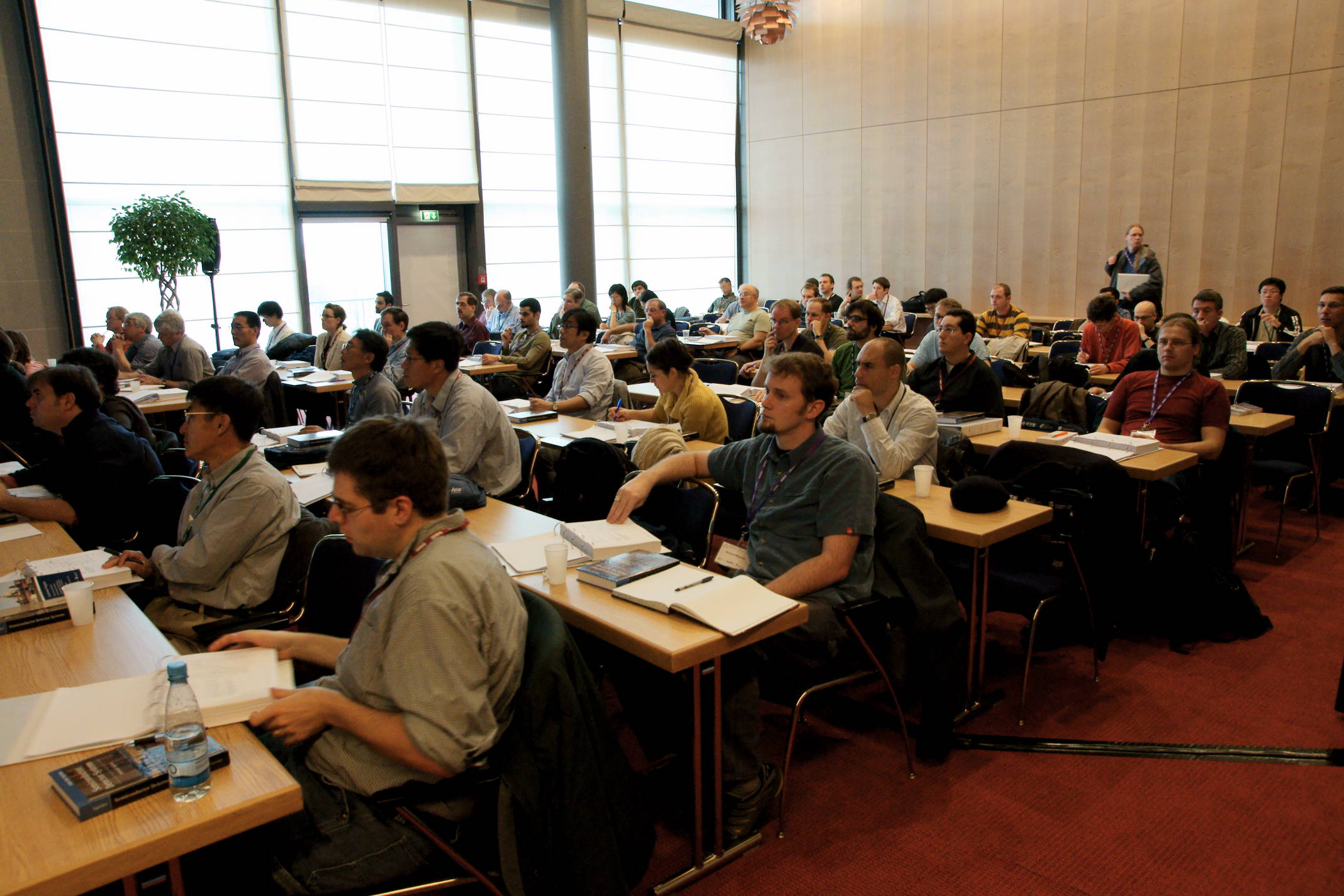
All courses are one day in length. Coffee and pastries will be available for participants of the short courses at 08:00, before the lectures begin at 08:30. Lunch, refreshments, lecture notes and a certification of completion are also provided as part of the registration fee.


Jennifer Huber
Short Course Co-Chair
LBNL, USA

Youngho Seo
Short Course Co-Chair
UCSF, USA

Stephen Derenzo
Short Course Co-Chair
LBNL, USA

Organizer: Gregg McKinney, Los Alamos National Laboratory, USA


Course Description

The MCNP Overview session will include four ~90-minute presentations and discussions on MCNP6 basics, physics, sources, and tallies (in this order). The Basics module will include distribution files, graphics software, execution, geometry input, plotting, and material input; the Physics module will include general physics input, low energy physics and related input, and high energy physics and related input; the Sources module will include standard source options and advanced source options (spontaneous decay, cosmic, background); and the Tallies module will include standard tally options and advanced tally options (tagging, Compton imaging, coincidence, ROC curves).


Instructor

Gregg McKinney is a senior Project Leader at Los Alamos National Laboratory (LANL). He received his M.S. and Ph.D. in Nuclear Engineering in 1984 and 1987 from the University of Washington. His principal work experience includes ~30 years of code development for Monte Carlo particle transport codes (MCNP, MCNPX), serving fifteen years as a code developer, four years as the MCNP Team Leader, four years as the MCNPX Team Leader, and six years as a Project Leader. He joined LANL in 1992. His experience in transport applications includes space systems, nuclear reactors, radiation therapy, and shielding. His current research interests include correlated particle production, fission physics, detector modeling, and coincidence counting.

Organizer: Paul O’Connor, Brookhaven National Laboratory, USA


Course Description

This one-day course is intended to introduce physicists and detector specialists to the fundamentals of integrated circuit front end design. The class begins with a discussion of low-noise signal processing and semiconductor devices and then delves into the details of implementing practical circuits in modern CMOS technology. A basic knowledge of detectors and electronics is assumed.


Course Outline

  1. Pulse Processing Fundamentals
    • Signal formation in detectors
    • Noise and gain mechanisms
    • Pulse processing for amplitude and timing extraction
  2. Semiconductor Technology for Integrated Circuit Front Ends
    • Operation and characteristics of MOS and bipolar transistors
    • Sub-micron CMOS and BICMOS technology
    • Feature size scaling
    • Radiation effects and reliability
    • Mixed-signal circuits
  3. Analog circuit design
    • The IC design process and CAD tools
    • Foundry access, multiproject services
    • Building blocks for the analog channel: charge-sensitive and pulse-shaping amplifiers, baseline stabilizers, peak detectors, track/hold, multiplexers, output stages
    • Analog-to-digital and time-to-digital converters (ADC and TDC)
  4. Packaging and Interconnect
  5. Application examples

Instructors

Paul O'Connor is associate Head of the Instrumentation Division at Brookhaven National Laboratory. After receiving the Ph.D. degree in solid-state physics from Brown University he worked from 1980-1990 at AT&T Bell Laboratories prior to joining BNL. His research interests are in the field of instrumentation systems for radiation detection, particularly low noise analog CMOS front-end circuits. He is author and co-author of about 80 publications and has been an IEEE member since 1980.

Christophe de La Taille is Technical Director of the French Institute for Particle Physics (IN2P3). After receiving an engineering and Ph.D. degree from Ecole Polytechnique, he joined CNRS/IN2P3 and worked on the readout of the ATLAS calorimeter at CERN/LHC and other high energy physics experiments. Prior to his present position, he was the leader of the electronics group at LAL, Orsay. His research interests are in the field of detectors and mixed signal ASIC design. He is author and co-author of about 168 publications and has been an IEEE member since 2003.

Veljko Radeka, Senior Scientist and Head of Instrumentation Division at Brookhaven National laboratory. His interests have been in scientific instruments, radiation detectors, noise and signal processing, and low noise electronics. He authored or coauthored about 190 publications. He is a Life Fellow of IEEE, a Fellow of APS, recipient of the 2009 Howard Wheeler Award from the IEEE and of the 2010 Radiation Instrumentation Outstanding Achievement Award, IEEE NPSS.

Organizer: Sara A. Pozzi, University of Michigan, USA


Course Description

The MCNPX-PoliMi code is a modified version of MCNPX v. 2.7.0 that provides unique capabilities for simulating correlated-particle measurements and detector response. This workshop will introduce new users to the capabilities of the MCNPX-PoliMi code and acquaint experienced users with new features.


Course Outline

  • MCNPX-PoliMi introduction
  • MCNPX-PoliMi source capabilities, including new models of fission
    • Spontaneous and neutron/gamma induced fission
    • Alpha-n sources
    • Combined sources such as MOX fuel
  • Detector-response simulations with the MPPost code
    • Organic scintillator (e.g. liquids and plastic)
    • Capture detectors (e.g. He-3)
    • Inorganic scintillators (e.g. NaI)
    • Special detectors (capture-gated)
  • Simulations of time-of-flight experiments
  • Simulations of multiplicity experiments

Instructors

Sara A. Pozzi is an Associate Professor in the Department of Nuclear and Radiological Sciences at the University of Michigan, where she established and is the leader of the Detection for Nuclear Nonproliferation Group. Her research interests include the development of new methods for nuclear materials detection, identification, and characterization for nuclear nonproliferation, nuclear material control and accountability, and national security programs. She has led many experimental campaigns on fissile material performed at national laboratories in the United States, in Italy, and in Russia. Prof. Pozzi’s research group is sponsored by the Department of Energy, Department of Homeland Security, Department of Defense, and other agencies. She is the co-author of the Monte Carlo code MCNPX-PoliMi, which is being used at over 50 institutions world-wide.

Her publication record includes over 300 papers in journals and international conference proceedings. She was invited to give over 50 seminars, both nationally and internationally. As the DNNG leader, she advises 14 doctoral students and many undergraduate students, and is the faculty advisor for the Institute of Nuclear Materials Management (INMM) student chapter.

Marek Flaska is an Associate Research Scientist in the Department of Nuclear and Radiological Sciences at the University of Michigan. During his Ph.D research, Dr. Flaska worked at the Institute for Reference Materials and Measurements in Geel, Belgium, the European Commission's Joint Research Center. He has many years of experience in research and development in the area of Monte Carlo simulations. His current field of interest is the development of new methods for accurate identification and characterization of special nuclear material and radioactive sources for applications in nuclear nonproliferation, nuclear safeguards, homeland security, and nuclear material control and accountability. He has performed numerous experiments and analyses using liquid and plastic scintillators, and capture-gated detectors.

Dr. Flaska co-mentored a number of undergraduate and graduate students and is the author or co-author of a large number of conference proceeding and journal papers.

Shaun D. Clarke is an Assistant Research Scientist in the Department of Nuclear and Radiological Sciences at the University of Michigan. He has more than 10 years of experience in Monte Carlo modeling and analysis, specifically with the codes MCNP4C, MCNP5, MCNPX and MCNP-PoliMi. Dr. Clarke also has several years of experience in radiation detection measurements. He has organized and performed international measurement campaigns involving measurements of special nuclear material with organic scintillation detectors and state-of-the-art digital electronics. His current research efforts are focused on Monte Carlo simulation techniques for active and passive detection systems for nuclear nonproliferation, safeguards, and homeland security applications. Dr. Clarke is author or coauthor of over 120 papers in conference proceedings and peer-reviewed journals.

Michael Hamel is a third year graduate student in the Department of Nuclear Engineering and Radiological Sciences at the University of Michigan. He has about five years of experience working with Monte Carlo simulations for his research projects including MCNP5, MCNPX and MCNP-PoliMi. Michael has also worked as a graduate student instructor several times for courses that feature the use of MCNP. Currently, Michael is working on developing spectroscopic capabilities for fast neutrons as part of a neutron and gamma ray imaging system for special nuclear material. He is the co-author of one journal article and has authored or co-authored nine conference papers.

Organizer: Woon-Seng Choong, Lawrence Berkeley National Laboratory, USA


Course Description

This 1-day course will discuss the photodetector technologies that are used for nuclear radiation detectors. The main photodetector used in scintillation spectroscopy at present is the photomultiplier tube (PMT) and its current status and on-going advances will be covered. The course will also present recent advances in silicon-based photodetectors such as unity gain silicon PIN diodes, drift detectors, high gain avalanche photodiodes (APDs), and the silicon photomultipliers (SiPMs). The potentials of wider-gap semiconductor-based photodetectors will be included. Front-end electronic readout designs for these different types of photodetectors will also be covered. Examples of detector configurations that employ various types of photodetectors in applications such as medical imaging and physics research will be given. Presentation materials will be provided as handouts. Some prior background in scintillation spectroscopy would be desirable but not essential.


Course Outline

  1. Vacuum-Based Photodetectors (PMT, MCP PMT, Hybrid Photodetectors)
  2. Solid State Photodetectors (Photodiode, APD, Geiger-Mode APD, SiPM)
  3. Signal Processing and Electronics
  4. Applications of Photodetectors in Physics Research and Nuclear Medicine

Instructors

Woon-Seng Choong is a Staff Scientist at the Lawrence Berkeley National Laboratory. He received a Ph.D in Physics from the University of California at Berkeley in 2000. His major research interests are in the development of novel and advanced instrumentation for biomedical imaging, specifically in nuclear medicine and image reconstruction. These include development of new scintillators for gamma ray detection, novel photodetectors, electronics for radionuclide imaging, and new detector designs and camera systems. Recently, he has been investigating advanced photodetector technologies such as multi-anode microchannel plate photomultiplier tubes and silicon photomultipliers.

Craig Woody is a Senior Physicist at the Brookhaven National Laboratory. He received his Ph.D. from the Johns Hopkins University in 1978. His research interests are primarily in the area of particle detectors and instrumentation for high energy and nuclear physics and medical imaging. These include various types of scintillating crystals and other types of scintillation detectors, optical readout devices and their associated electronics, laser systems, and gas detectors for particle tracking and imaging applications. Other primary research interests are in relativistic heavy ion physics with the PHENIX Experiment at the Relativistic Heavy Ion Collider at Brookhaven.

Mitch Newcomer is currently director of the Penn Instrumentation group. He received a BA in Physics from Penn in 1976 and has remained at Penn since. Over the past 35+ years he has specialized in front end electronics for Nuclear, High Energy, and Medical Physics. This includes the Front End electronics for the Kamiokande II and SNO large water Cherenkov detectors, for the TOF system and Central Open Tracker readout of the upgraded CDF detector (1999), and the analog signal processing for the 340K channel ATLAS Transition Radiation Tracker. Currently he has primary responsibility for the design of the analog signal processing ASIC for the upgraded ATLAS EM Calorimeter. In medical physics applications he has re-designed the front end electronics of a PMT based commercial whole body PET scanner to add a TOF capability that has realized a precision 370ps FWHM, as well as design the readout system for a 16 plane pixelized, micromegas proton therapy dose imager.

Anton Tremsin is a Full Research Physicist at the Space Sciences Laboratory of University of California at Berkeley. He has received his degrees in Physics in 1992 from Moscow Institute of Physics and Technology and the Russian Academy of Sciences and continued as a Royal Society Postdoctoral Fellow at Leicester University, UK. In his research at UC Berkeley Dr. Tremsin is working on the development of new technologies for particle detection (photon/electron/ion/neutron) with high spatial and temporal resolution based on microchannel plate technology. His current interests are in the development of vacuum detectors with efficient UV and soft X-ray photocathodes, fast readout electronics capable of event counting at high counting rates and in the development of novel neutron and electron counting detectors for beamline applications at spallation neutron sources and synchrotrons. These new detectors enable novel non-destructive studies in materials sciences, geophysics, biomedical imaging, energy research and industrial applications.

Organizer: Steve Derenzo, Lawrence Berkeley National Laboratory, USA

Course Description

This one-day course will cover the basic concepts that describe the randomness in measured data, how to find the most likely model that fits the data, and how to determine the statistical variability of the model parameters


Course Outline

  1. Random distributions
    • Poisson
    • Gaussian
    • Moments
    • Error propagation
    • Monte Carlo simulation
  2. Parameter estimation
    • Chi-squared
    • Maximum likelihood
    • Iterative methods
  3. Statistics of radiation detectors
    • Energy resolution
    • Position resolution
    • Timing resolution
    • Minimum detectable signals
    • Paralyzing and non-paralyzing dead time
  4. Statistics in image reconstruction
    • Analytical and iterative image reconstruction methods
    • Applications to emission and transmission tomography
    • Image variance
    • Cramer-Rao lower limits on the image variance

Instructors

Stephen Derenzo is a Senior Scientist at the Lawrence Berkeley National Laboratory and Adjunct Professor in the Electrical Engineering and Computer Science Department at UC Berkeley. Statistics and data analysis are essential tools of his research and are also important components of the courses he teaches at UC Berkeley. For the past 24 years he has lead a search for new heavy scintillators and currently heads a project for the discovery of scintillation detector materials that uses automation to increase the rate of synthesis and characterization. He recently completed a comprehensive analysis of the statistical limitations of timing precision in scintillation detectors. He has authored or co-authored over 200 technical publications, seven patents, and one textbook. He has received two awards from the IEEE Nuclear and Plasma Sciences Society: the Merit Award in 1992 and the Radiation Instrumentation Outstanding Achievement Award in 2001. He became an IEEE Fellow in 2000.

Ana Kupresanin received her Ph.D. in statistics at Arizona State University in 2008. Her research is in functional data analysis and uncertainty quantification in engineering applications. She is currently a Statistician at the Lawrence Livermore Laboratory where her work includes statistical problems related to the stockpile stewardship program. She has mentored several Ph.D. students during their LLNL internships and maintains a research collaboration with UC Davis. She coauthored the book Statistical Computing in C++ and R.

Michel Defrise received the Ph.D. degree in theoretical physics from the University of Brussels in 1981, and was a visiting professor in the Department of Radiology of the University of Geneva in 1992-1993. He is currently research professor in the Department of Nuclear Medicine of the Vrije Universiteit Brussel (Belgium). His research interests include 3-D image reconstruction in nuclear medicine (PET and SPECT) and in CT. He is Fellow of the IEEE and of the Institute of Physics, and was a recipient in 2011 of the Edward J Hoffman Medical Imaging Senior Scientist Award of the IEEE and of the Edward J Hoffman Memorial Award of the Society of Nuclear Medicine.

Organizer: Peder Larson, University of California, San Francisco, USA


Course Description

Magnetic resonance imaging (MRI) is a powerful modality, providing a variety of anatomical and functional contrast for soft tissues in arbitrarily oriented 2D planes and 3D volumes. This is achieved by manipulating the magnetic moments of nuclear spins in a magnetic field using magnetic field gradients and radiofrequency waves. MRI can provide contrast based on magnetic properties (T1, T2), proton density, flow, perfusion, diffusion, stiffness, blood oxygenation (functional MRI), and metabolism (MR spectroscopy).

This short course aims to teach the basic principles behind magnetic resonance imaging (MRI). The topics taught will include: physics of magnetic resonance, including resonance, excitation, and relaxation; image formation with excitation pulses and gradients fields; image reconstruction via the Fourier Transform; MRI scanner hardware, including magnets and coils; image contrast mechanisms; artifacts due to flow, motion, and field variations; and in vivo applications.


Instructors

Peder Larson is an Assistant Professor in the Department of Radiology and Biomedical Imaging at the University of California, San Francisco, where he teaches a graduate course on “Principles of MRI”. He received his PhD in Electrical Engineering from Stanford University in 2007, followed by a postdoctoral fellowship in the Department of Radiology at UCSF. He is an IEEE member and has co-authored 38 peer-reviewed papers. Dr. Larson's research program is centered on MR pulse sequence development, including MR physics, RF pulse design, acquisition strategies, and reconstruction techniques. Specifically, his interests are in methods development for MRI with hyperpolarized carbon-13 agents, a promising new metabolic imaging modality, semi-solid tissue MRI for positive contrast of tissues such as tendons, bone, and myelin that a re invisible with conventional MRI techniques.

Vasily Yarnykh, PhD, is an Associate Professor in the Department of Radiology at the University of Washington (Seattle, WA). Dr. Yarnykh became involved in magnetic resonance research during his undergraduate and doctorate studies at the Lomonosov Moscow State University (Moscow, Russia). After post-doctoral fellowship at the University of Washington, he has been continuing his research career as a faculty member. Dr. Yarnykh has authored and co-authored 54 peer-reviewed journal articles and 4 US patents. Dr. Yarnykh’s research has been focused on the quantitative imaging methodology and development of new pulse sequences primarily for neuroimaging and vascular applications.

Niranjan Balu is currently a faculty of the Department of Radiology, University of Washington, Seattle serving in the position of Acting Instructor. He received his PhD in Biomedical Engineering from the University of Washington, Seattle. Following a post-doctoral position at the Vascular Imaging Lab at the University of Washington specializing in vascular MRI, he is currently working on developing pulse sequences for cardiovascular MRI of multiple vascular beds. Dr. Balu has authored and co-authored 21 peer reviewed publications in the field of vascular MRI. His specific research interests are in developing pulse sequences and reconstruction methods for diagnosis of cardiovascular pathology.

Organizer: Lars Furenlid, University of Arizona, USA


Course Description

Multimodality imaging systems are used increasingly in clinical medicine in an attempt to get better diagnostic or scientific information by acquiring images depicting different aspects of the object, such as physiological and functional characteristics. A newly emerging methodology with similar goals is adaptive imaging in which an initial image of a particular subject is acquired and then used to modify the data-acquisition hardware or protocol for obtaining a second image from the same or a different modality. In this case the imaging process is necessarily nonlinear because the characteristics of the second system depend on the object being imaged.

Because the goal of both adaptive and multimodality imaging is to obtain better information about a patient, the proper measure of image quality assesses how well this information can be extracted from the whole set of image data by a relevant observer. This approach, known as objective or task-based assessment of image quality, is well developed for single modalities and for linear, object-independent systems, but little has been done on applying it to adaptive and multimodality systems.

This course will review the basic principles of task-based assessment of image quality and discuss how they can be applied to adaptive and multimodality systems. It will cover the basic theory, hardware implementations, computational requirements and clinical applications.


Course Outline

  • Overview of multimodality imaging systems
  • Introduction to adaptive imaging
  • Principles of task-based assessment of image quality
  • Image quality and dose considerations
  • Task-based analysis of adaptive and multimodality systems
  • Comparisons between hardware designs
  • Data-analysis methods and computational requirements
  • Applications

Instructors

Lars Furenlid was educated at the University of Arizona and the Georgia Institute of Technology. He is currently a Professor at the University of Arizona and associate director of the Center for Gamma-ray Imaging, with appointments in the Department of Radiology, the College of Optical Sciences, the Graduate Interdisciplinary Degree Program in Biomedical Engineering, and the Arizona Cancer Center. He was a staff scientist at the National Synchrotron Light Source at Brookhaven National Laboratory. His major research area is the development and application of detectors, electronics, and systems for biomedical imaging. He was awarded the 2013 IEEE Radiation Instrumentation Outstanding Achievement Award.

Matthew A. Kupinski is a Professor at The University of Arizona with appointments in the College of Optical Sciences, the Department of Medical Imaging, and the program in Applied Mathematics. He performs theoretical research in the field of image science. His recent research emphasis is on quantifying the quality of multimodality and adaptive medical imaging systems using objective, task-based measures of image quality. He has a BS in physics from Trinity University in San Antonio, Texas, and received his PhD in 2000 from the University of Chicago. He is the recipient of the 2007 Mark Tetalman Award given out by the Society of Nuclear Medicine and is a member of the OSA and SPIE. Contact him at College of Optical Sciences, The University of Arizona, 1630 E. University Blvd., Tucson, Arizona 85721; mkupinski@optics.arizona.edu

Harrison Barrett Dr. Barrett received a bachelor's degree in physics from Virginia Polytechnic Institute in 1960, a master's degree in physics from MIT in 1962, and a Ph.D. in applied physics from Harvard in 1969. He worked for the Raytheon Research Division until 1974, when he came to the University of Arizona. He is a Regents Professor in the College of Optical Sciences and the Department of Medical Imaging in the College of Medicine, and he has appointments in Applied Mathematics, Biomedical Engineering and the Arizona Cancer Center. He is a fellow of the Optical Society of America, the Institute of Electrical and Electronic Engineers, the American Physical Society and the American Institute of Medical and Biological Engineering.

Dr. Barrett has obtained 25 U. S. patents and written over 200 technical papers, and 60 students have received Ph. D. degrees under his direction. In collaboration with Kyle J. Myers, he has written a book entitled Foundations of Image Science, which in 2006 was awarded the First Biennial J. W. Goodman Book Writing Award from OSA and SPIE.

His other awards include a Humboldt Prize, the 2000 IEEE Medical Imaging Scientist Award, an E. T. S. Walton Award from Science Foundation Ireland, the 2005 C. E. K. Mees Medal from the Optical Society of America and an honorary doctorate from the University of Ghent in Belgium. He was the 2011 recipient of the IEEE Medal for Innovations in Healthcare Technology and also the 2011 recipient of the SPIE Gold Medal of the Society. In 2014 he was elected to the National Academy of Engineering, and he received the Paul C. Aebersold Award of the Society of Nuclear Medicine and Molecular Imaging.

Organizer: Anatoly Rosenfeld, University of Wollongong , Australia


Course Description

This one day course provides a broad overview of the principles and physics of modern radiation therapy for cancer, including X-ray and charged particles external beam radiotherapy, brachytherapy, and applicable radiation dosimetry. Such a course naturally cannot cover each modality in detail, but rather aims to provide an outline of, and rationale behind each radiotherapy technique. The latest advancements in the field and the underlying physics will be described. The course will cover the principles of radiation dosimetry, quality assurance required for contemporary radiation therapy, and required radiation detectors. Semiconductor radiation detectors will be covered in greater detail.

The course will benefit electronic engineers and physicists who want to learn about state-of-the–art or refresh their knowledge of a fast growing field of radiotherapy technology. It will be equally useful to radiation detector instrumentation scientists who will get new ideas on developing detectors required for quality assurance and dose imaging in radiation therapy. Students attending the course will see new opportunities for their future research and employment.


Course Outline

  1. X-ray External Beam Radiation Therapy
    • Principles of radiotherapy (including radiobiological considerations)
    • 3D Conformal Radiation Therapy, Intensity Modulated Radiation Therapy / Volumetric Modulated Arc Therapy and optimization techniques
    • Stereotactic Radiosurgery and Radiotherapy
    • Modern linear accelerators
    • Clinical applications
  2. Brachytherapy
    • Principles of brachytherapy (including radiobiology)
    • High- and Low- Dose Rate brachytherapy
    • Clinical applications
  3. Charged particle radiotherapy
    • Short history of particle therapy
    • Current and future accelerators for proton and ion therapy
    • Radiobiology of protons and heavier ions
    • Treatment planning: what is different compared to photons
    • Clinical applications: past, present and future
  4. Radiation dosimetry
    • Principles of radiation dosimetry
    • Micro-and nano-dosimetry basics
    • Semiconductor dosimetry for quality assurance in X-ray, neutron and charged particle radiotherapy

Instructors

Anatoly Rosenfeld is a Professor of Medical Physics and Director of Centre for Medical Radiation Physics (CMRP) at University of Wollongong, Australia. His research interest is in the field of semiconductor radiation detectors for dosimetry and microdosimetry and its applications for real time quality assurance in conventional and hadron radiotherapy, space and avionics. Prof Rozenfeld attracted many competitive grants as PI and published more than 200 peer-reviewed articles, book chapters and holds numerous patents on radiation instruments. He has earned a MSc (with distinction) and PhD from Leningrad Politechnic Institute (Russia) and Institute for Nuclear Research (Ukraine) respectively.

Vladimir Feygelman earned his PhD in Physical Chemistry in the former USSR and was active in the field of Photochemistry. He is currently an Associate Member of the Department of Radiation Oncology at Moffitt Cancer Center in Tampa, Florida. He is an experienced ABR-certified clinical physicist, with research interests centered on radiation dosimetry and quality assurance of complicated dynamic treatments. This includes evaluation and improvement of dosimetry systems, particularly diode arrays, clinical comparisons, applications to different dose modulation techniques, and 4D dosimetry. Over the span of his career, Dr. Feygelman has over 50 peer-reviewed publications.

Marco Zaider is a Professor and Attending and Head of Brachytherapy Physics at Memorial Sloan Kettering Cancer Center, NY, USA, and Professor of Physics in Radiology at Weill Cornell Medical College. He received his PhD in nuclear physics at Tel Aviv University and, following a post-doctoral fellowship at Los Alamos National Laboratory, has remained actively involved in medical physics, microdosimetry and biomathematics. Prof. Zaider is the author (with Harald Rossi) of two textbooks and numerous book chapters and articles. He is the recipient of the 2007 Franz Edelman Award for Achievement in Operations Research and a Fellow of the American Association of Physicists in Medicine.

Reinhard Schulte is a Professor of Radiation Medicine in the School of Medicine of Loma Linda University, and works as Translational Research Specialist in the James M. Slater Proton Treatment and Research Center, Department of Radiation Medicine, Loma Linda University Medical Center. He received his Diploma in Physics from Dortmund University, Germany in 1978 and his Doctorate in Medicine (Dr. med., summa cum laude) from the University of Cologne, Germany in 1986. He is Principal Investigator on an NIH-funded project to develop proton CT and is involved in two large European Research Consortia related to proton therapy. Dr. Schulte also has more than 20 years of experience in clinical proton therapy and is board certified radiation oncologist in the United States and Germany.

Organizers: Harry Tsoumpas, University of Leeds, United Kingdom & Kris Thielemans, UCL, United Kingdom


Course Description

The growing success of PET and SPECT imaging combined with CT or MR has led to the evolution of molecular imaging modalities that assist in improving diagnosis and staging of diseases. Emerging PET and SPECT imaging is used to drive patient therapy, therefore reliable image quantification and high image quality are important factors. Image reconstruction methods have a key role in converting the measurement to a meaningful image and along with new hardware developments offer a stimulating research environment for advancements of PET and SPECT imaging.

This course will provide an overview of iterative tomographic image reconstruction methods. It will start with fundamentals of image reconstruction for PET and SPECT imaging. It will then describe current methods to account for the complicated physics of the acquisition process and other factors such as motion. The third part of the course will cover demonstrations and practical exercises with an open source library for tomographic image reconstruction (STIR, stir.sf.net). The attendees will have the opportunity to simulate and reconstruct data, perform scatter correction, motion correction and other tasks. Prerequisite knowledge includes basic principles of physics of γ-rays, statistics, calculus, and elementary linear algebra. For the practical sessions, students are encouraged to bring their own laptop and install STIR before attending the course. Basic knowledge of linux terminal commands is recommended. Knowledge of C++ is not necessary but will be helpful.


Course Outline

  • Tomographic reconstruction introduction
  • Basics of fully 3D iterative reconstruction for PET and SPECT
  • Basics of model-based image reconstruction (e.g. Scatter, Motion, PSF, Anatomical priors)
  • Advanced iterative reconstruction (e.g. TOF, MLAA, 4D)
  • Demos and practical exercises with open source software

Instructors

Johan Nuyts is a professor of the Faculty of Medicine at KU Leuven, Belgium. He is with the Department of Nuclear Medicine and with the Medical Imaging Research Center (MIRC). He received his Ph.D. in applied sciences from KU Leuven in 1991, on the subject of image reconstruction and quantification in SPECT. He co-authored about 120 scientific journal papers. His main research interest is in iterative reconstruction in PET, SPECT and CT. Ongoing research projects focus on maximum-a-posteriori reconstruction in emission tomography, iterative reconstruction in CT and tomosynthesis, attenuation correction in PET/CT, PET/MRI and TOF-PET, and motion correction in PET and CT.

Kris Thielemans is a Senior Lecturer at University College London (UCL) and is an IEEE senior member. He received his Ph.D degree in String Theory from KU Leuven in 1994. Prior to UCL, he has been working as a Researcher at Hammersmith (London, UK) for the Medical Research Council and General Electric, and at King’s College London (KCL). His research interests encompass all aspects of quantitative PET image reconstruction with emphasis on the development of advanced reconstruction techniques for PET and SPECT including motion correction. He developed along with others an open source software for tomographic image reconstruction (STIR) which has been cited more than 200 times.

Charalampos Tsoumpas is a Lecturer of Medical Imaging at the University of Leeds and Honorary Research Officer with the Leeds Teaching Hospitals since 2013. He received his Ph.D. degree in Parametric Image Reconstruction from Imperial College London in 2008 and worked as a post-doctoral fellow at KCL on PET-MR. He is a Senior Member of IEEE and Fellow of Higher Education Academy. He has contributions in more than 30 peer-reviewed papers, 20 IEEE conference records and two patents with GE Healthcare. His research interests include statistical image reconstruction and acquisition system modelling for more accurate and precise PET and PET-MR imaging.

Short Course Schedule

Date Room 618-619 Room 616-617 Room 602-604
Saturday 8 November 2014 SC1 SC2  
Sunday 9 November 2014 SC3 SC5 SC4
Monday 10 November 2014 SC6 SC7  
Tuesday 11 November 2014 SC8 SC9